©The Author(s) 2020.
World J Clin Cases. Jun 6, 2020; 8(11): 2359-2363
Published online Jun 6, 2020. doi: 10.12998/wjcc.v8.i11.2359
Published online Jun 6, 2020. doi: 10.12998/wjcc.v8.i11.2359
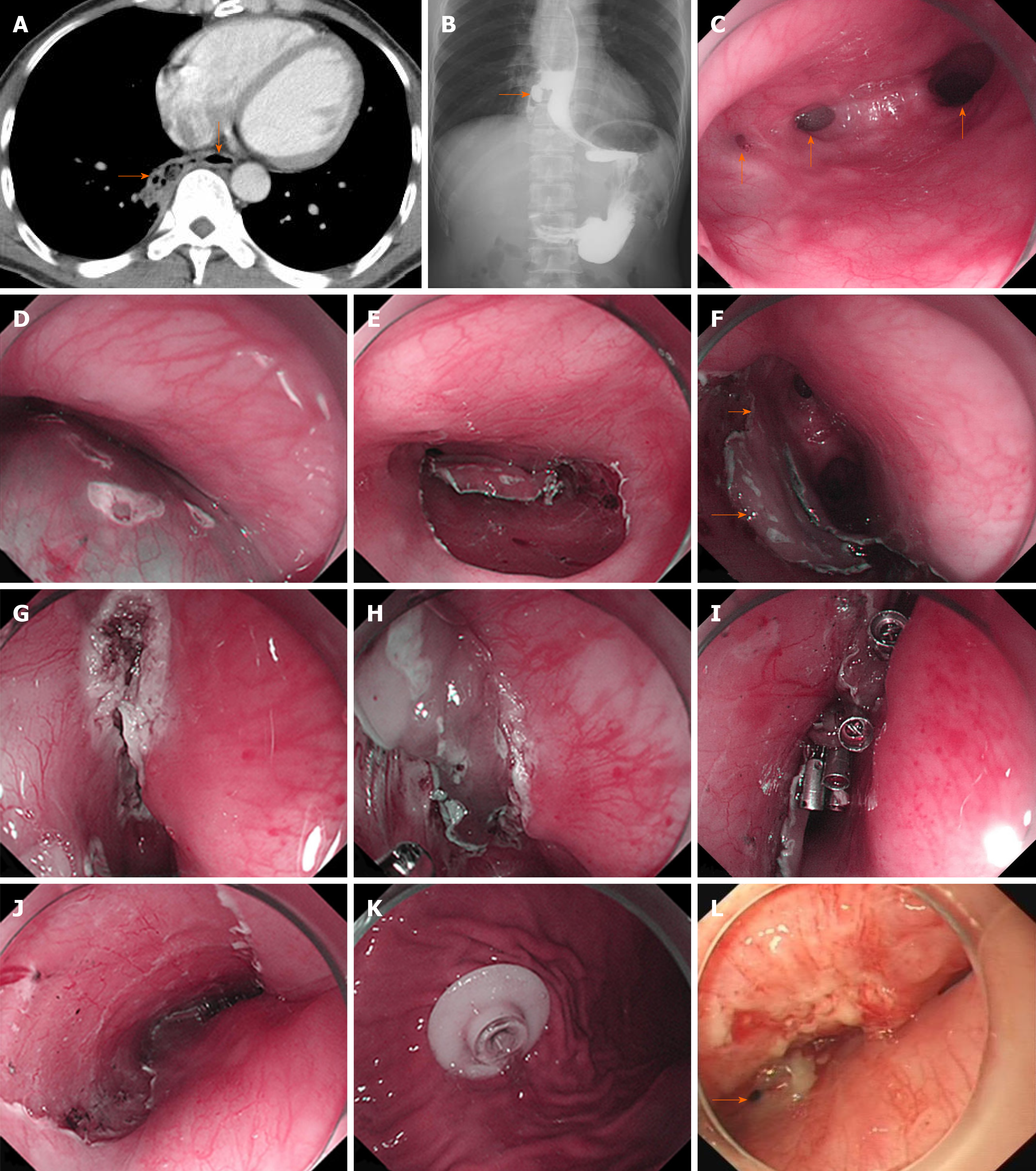
Figure 1 Results of examinations and treatment.
A: Computed tomography scan (rightward arrow: Fistula; downward arrow: Esophageal lumen); B: Esophagram revealed the presence of esophageal fistula (arrow) around the 9th vertebra level; C: Endoscopic examination confirmed the presence of two fistulas, which are 0.3 cm (left arrow) and 0.5 cm (middle arrow) in diameter and the esophageal lumen (right arrow); D and E: Submucosal injection was followed by (E) endoscopic submucosal dissection; F: The mucosal flap (lower arrow) with the caudal edge reserved as the pedicle (upper arrow) was made ready after endoscopic submucosal dissection; G and H: Ablation of the surrounding mucosa before (H) covering the fistulas with the flap; I: Several titanium clips were used for fixation; J: Ulcer area left after endoscopic submucosal dissection; K: Percutaneous gastrostomy; L: Forty-five days after the endoscopic surgery, the larger fistula (arrow) reduced from 0.5 cm to 0.2 cm and the smaller one was closed.
- Citation: Zhang YH, Du J, Li CH, Hu B. Endoscopic pedicle flap grafting in the treatment of esophageal fistulas: A case report. World J Clin Cases 2020; 8(11): 2359-2363
- URL: https://www.wjgnet.com/2307-8960/full/v8/i11/2359.htm
- DOI: https://dx.doi.org/10.12998/wjcc.v8.i11.2359


