©The Author(s) 2019.
World J Clin Cases. Dec 26, 2019; 7(24): 4327-4333
Published online Dec 26, 2019. doi: 10.12998/wjcc.v7.i24.4327
Published online Dec 26, 2019. doi: 10.12998/wjcc.v7.i24.4327
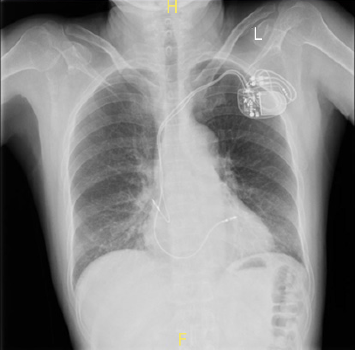
Figure 1 Posteroanterior chest X-ray shows the tip of the atrial lead located outside the cardiac shadow.
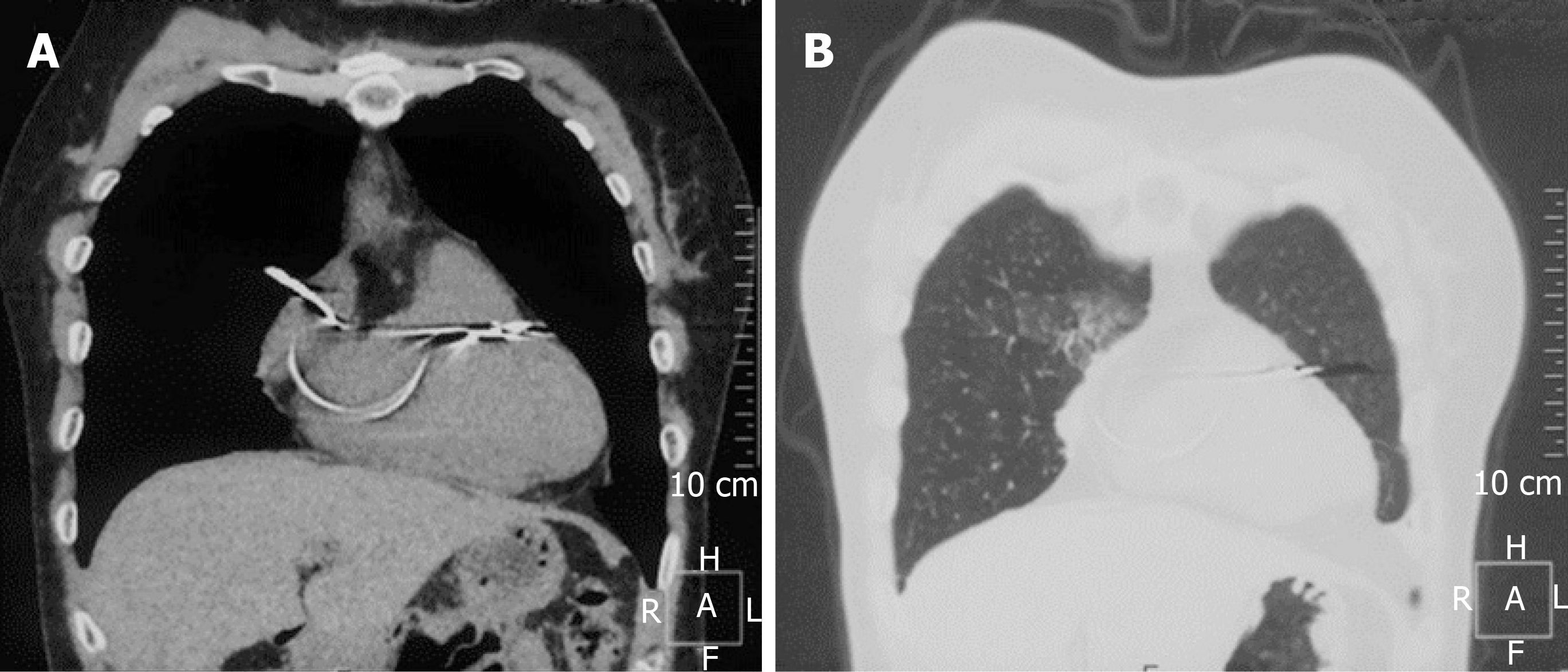
Figure 2 The chest computed tomography shows the atrial lead tip migrating into the lung tissue with signs of local inflammation.
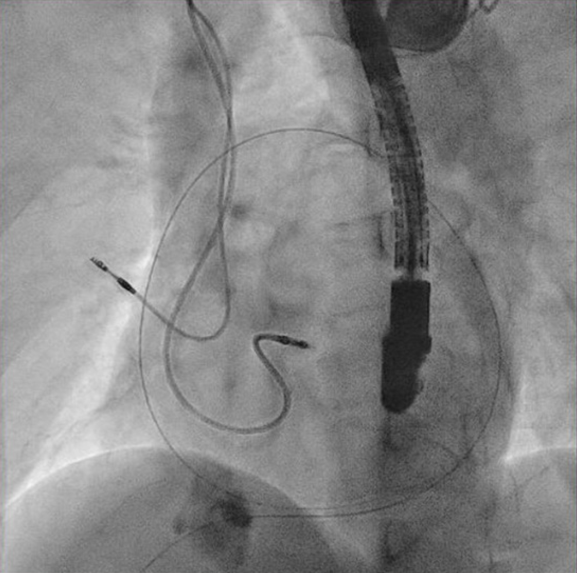
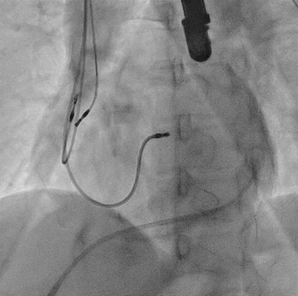
Figure 3 After percutaneous subxiphoid pericardial puncture, a soft floppy-tipped guidewire was advanced along the lateral heart border in the left anterior oblique projection.
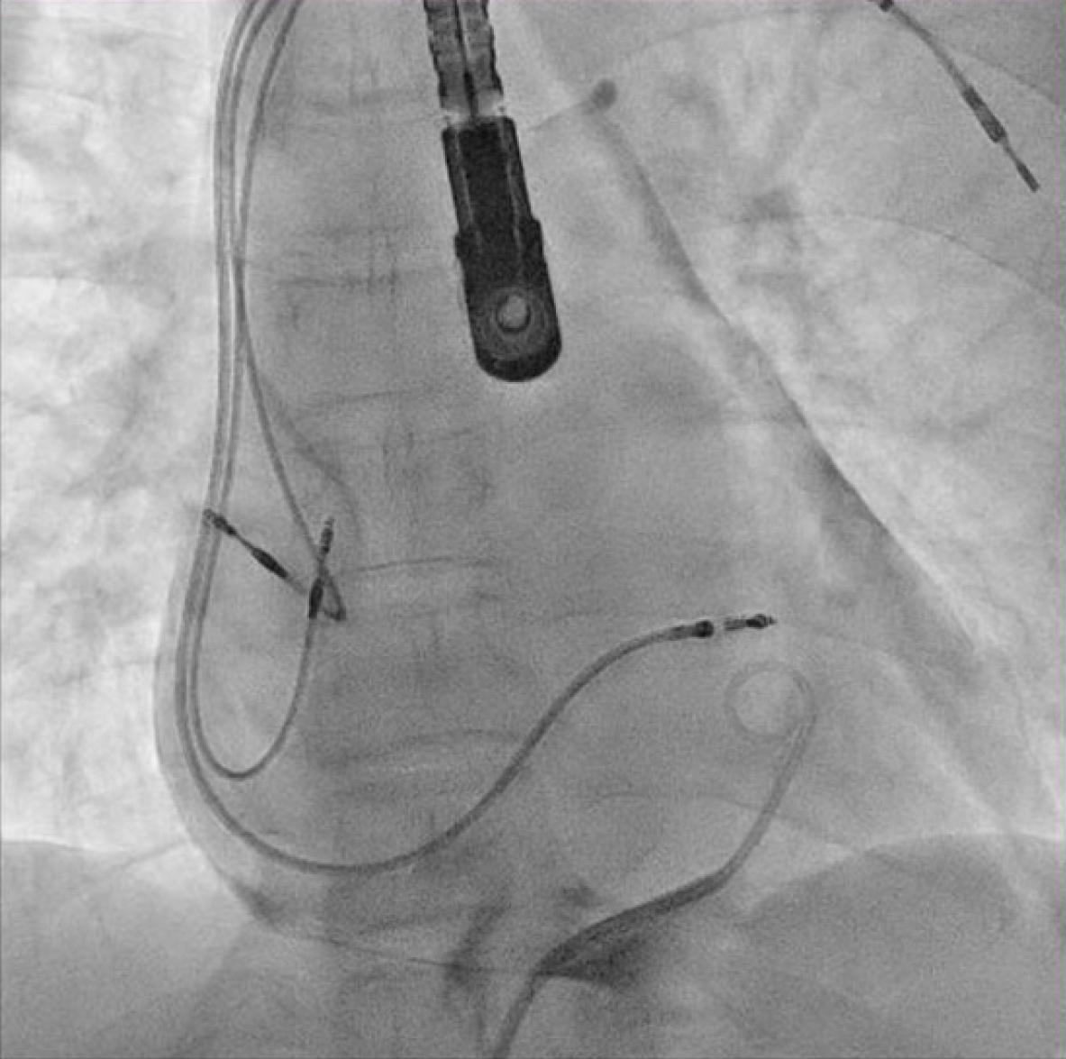
Figure 4 A new active fixation atrial lead was implanted at a different site with less tension, prior to removal of the culprit lead.
Figure 5 The culprit atrial lead was removed with simple traction after the active screw was retracted.
- Citation: Zhou X, Ze F, Li D, Li XB. Percutaneous management of atrium and lung perforation: A case report. World J Clin Cases 2019; 7(24): 4327-4333
- URL: https://www.wjgnet.com/2307-8960/full/v7/i24/4327.htm
- DOI: https://dx.doi.org/10.12998/wjcc.v7.i24.4327


