Copyright
©The Author(s) 2019.
World J Clin Cases. Nov 26, 2019; 7(22): 3821-3831
Published online Nov 26, 2019. doi: 10.12998/wjcc.v7.i22.3821
Published online Nov 26, 2019. doi: 10.12998/wjcc.v7.i22.3821
Figure 1 Images of our patient with allergic fungal rhinosinusitis accompanied by allergic bronchopulmonary aspergillosis.
A: Coronal CT scan of the sinuses revealed a ground-glass opacity filling the right maxillary and ethmoid sinuses, along with bone absorption in the medial wall of the right maxillary sinus; B: Axial CT scan of the sinuses; C: A well-defined mass, located in the right maxillary and ethmoid sinuses and displaying obvious hypointense features, was observed on coronal T2-weighted image; D: There was a peripheral enhancement of the mass on gadolinium-enhanced axial T1-weighted image.
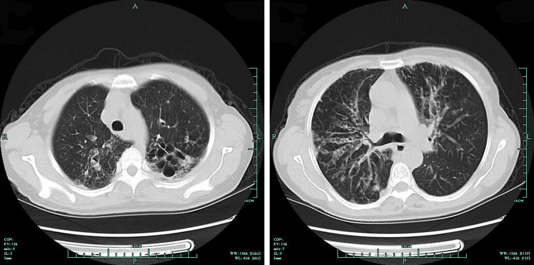
Figure 2 Chest CT revealed columned and cystiform bronchiectasis in the bilateral bronchus, surrounded by a large number of spotted and funicular high-density lesions.
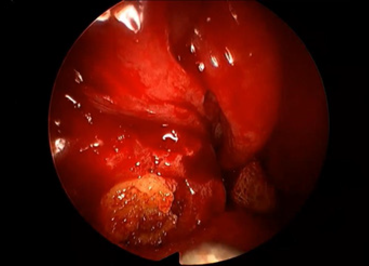
Figure 3 A large amount of yellow, jam-like mucin in the right maxillary and ethmoid sinuses during the surgery.
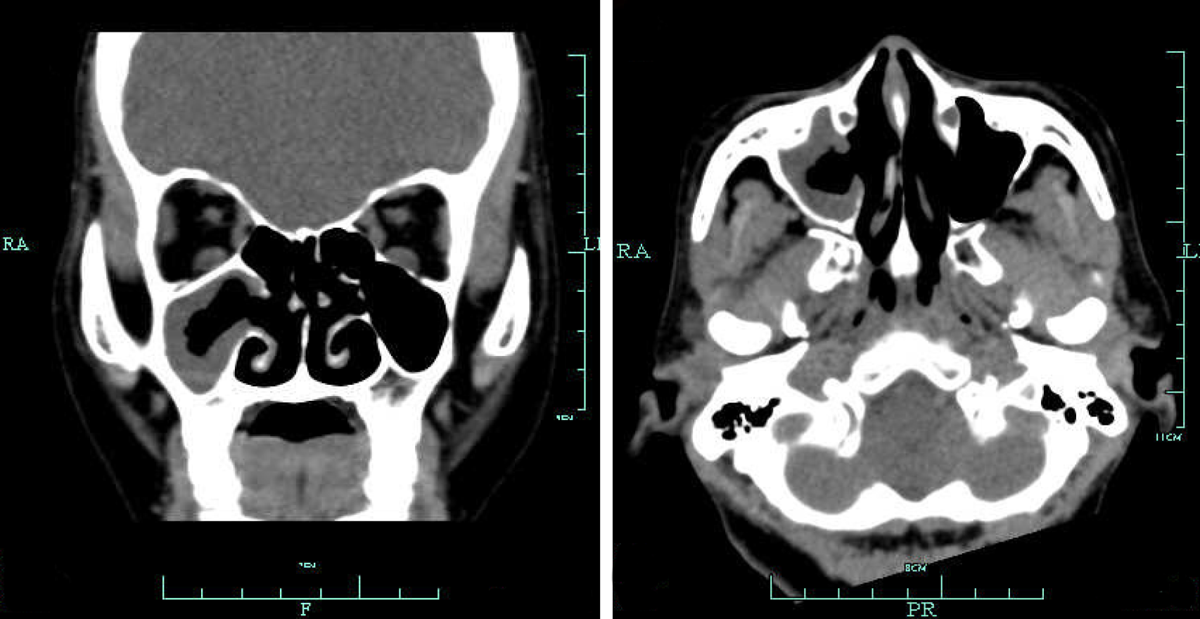
Figure 4 Sinus CT images indicating a good recovery, with secretions in the right maxillary sinus after surgery.
- Citation: Cheng KJ, Zhou ML, Liu YC, Zhou SH. Allergic fungal rhinosinusitis accompanied by allergic bronchopulmonary aspergillosis: A case report and literature review. World J Clin Cases 2019; 7(22): 3821-3831
- URL: https://www.wjgnet.com/2307-8960/full/v7/i22/3821.htm
- DOI: https://dx.doi.org/10.12998/wjcc.v7.i22.3821


