©The Author(s) 2019.
World Journal of Clinical Cases. Sep 6, 2019; 7(17): 2549-2555
Published online Sep 6, 2019. doi: 10.12998/wjcc.v7.i17.2549
Published online Sep 6, 2019. doi: 10.12998/wjcc.v7.i17.2549
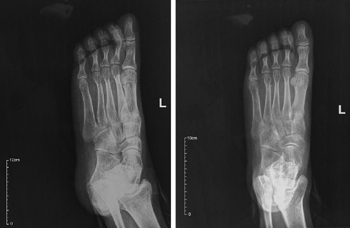
Figure 1 Plain radiography of the patient’s left plantar.
Both of the front view and foot oblique view show nothing abnormal in bones.
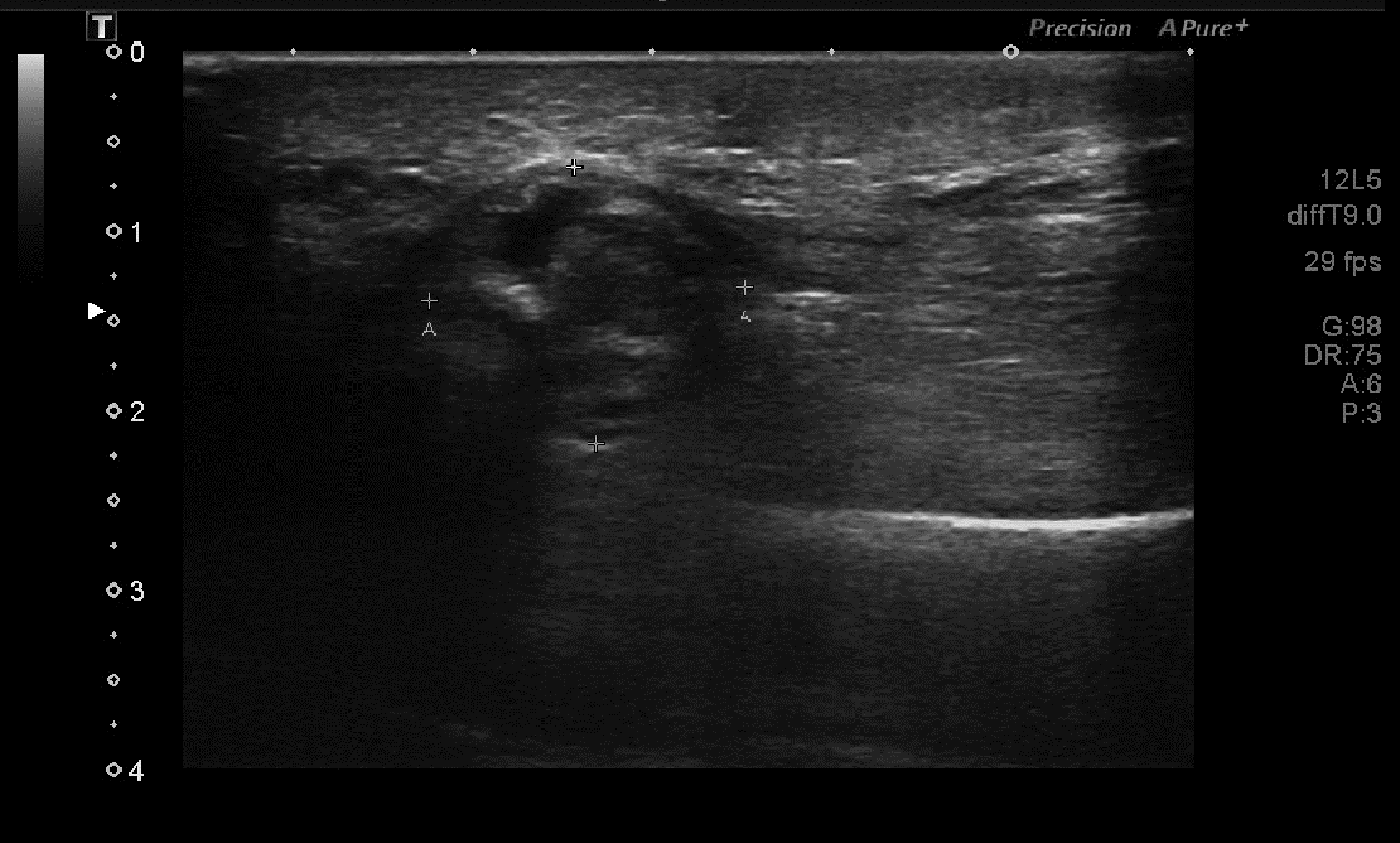
Figure 2 Doppler ultrasonography showed an inhomogeneous mass in the plantar area of the left foot.
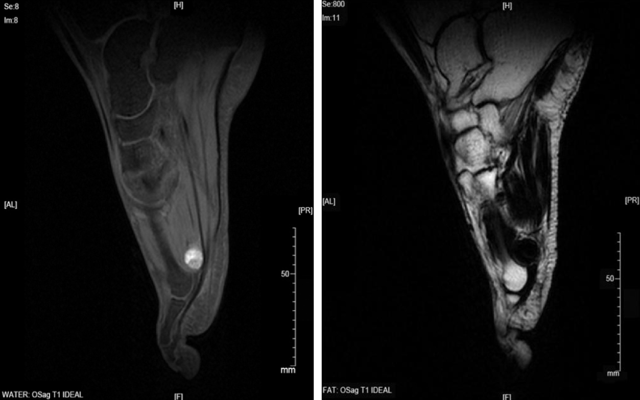
Figure 3 Magnetic resonance imaging suggested that the lesion could be a benign tumor or tumor-like lesion, such as inflammatory granulation or a giant cell tumor in a tendon sheath.
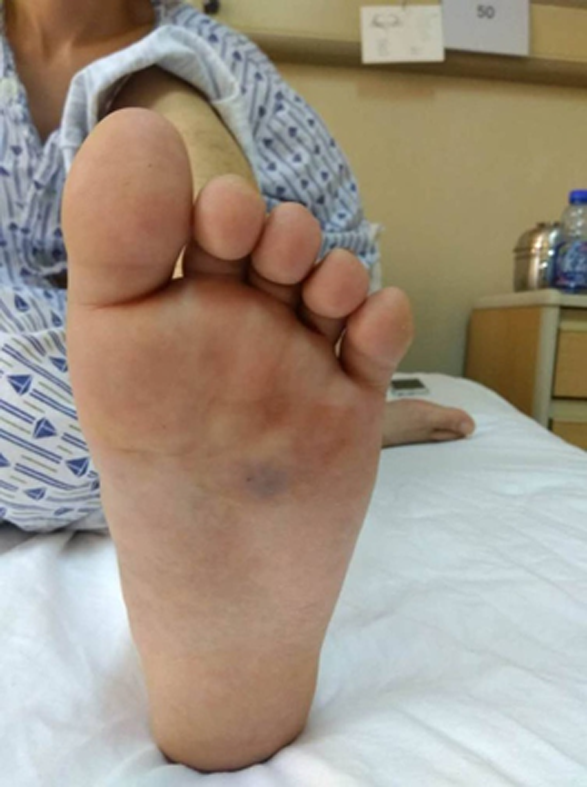
Figure 4 Physical examination showed a swelling in the left plantar area and ecchymosis (1.
5 cm2 × 1.5 cm2) in the left plantar region with accompanying tenderness.
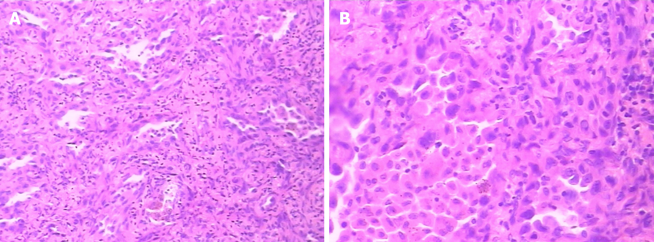
Figure 5 Hematoxylin-eosin staining showed a sign of tumor cell type.
A: Magnification is 100 times; B: Magnification is 200 times.
- Citation: Gao J, Yuan YS, Liu T, Lv HR, Xu HL. Synovial sarcoma in the plantar region: A case report and literature review. World Journal of Clinical Cases 2019; 7(17): 2549-2555
- URL: https://www.wjgnet.com/2307-8960/full/v7/i17/2549.htm
- DOI: https://dx.doi.org/10.12998/wjcc.v7.i17.2549


