Copyright
©The Author(s) 2019.
World J Clin Cases. Jun 6, 2019; 7(11): 1344-1350
Published online Jun 6, 2019. doi: 10.12998/wjcc.v7.i11.1344
Published online Jun 6, 2019. doi: 10.12998/wjcc.v7.i11.1344
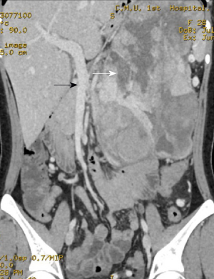
Figure 1 Preoperative computed tomography.
Preoperative computed tomography shows a huge cystic-solid mass in the left upper abdomen, which compressed the superior mesenteric vein (Black arrow); necrosis can be seen in the mass (White arrow).
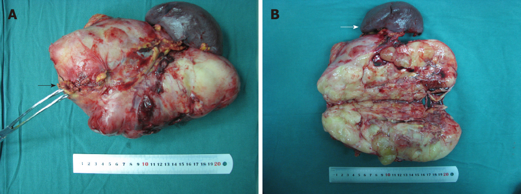
Figure 2 Surgical procedure.
The pancreatic tail was removed. A: The giant mass in the pancreatic tail was approximately 28.0 cm × 19.0 cm × 8.0 cm. The black arrow shows the normal pancreatic tissue; B: The mass was longitudinally opened. The gray-white and fish-like incisal surface can be seen. The spleen is shown by the white arrow.
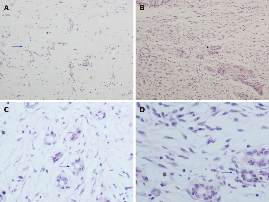
Figure 3 Histopathology.
A: Coexisting areas of well-differentiated liposarcoma and the pancreatic canal (H and E × 100); B: Coexisting areas of dedifferentiated liposarcoma and the pancreatic canal (H and E × 100); C: Coexisting areas of well-differentiated liposarcoma and the pancreatic canal (H and E × 400); D: Coexisting areas of dedifferentiated liposarcoma and the pancreatic canal (H and E × 400). Blue arrows show areas containing the pancreatic canal. H and E: Hematoxylin and eosin.
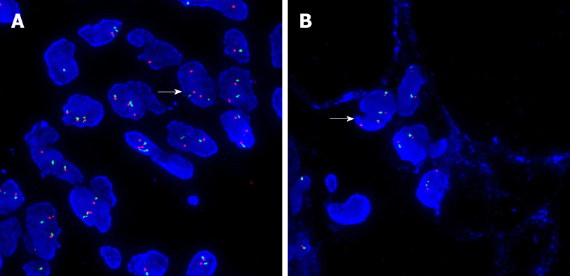
Figure 4 Fluorescence in situ hybridization results.
A: MDM2 gene amplification of the mass was positive (White arrow; as shown by three red dots and two green dots); B: MDM2 gene amplification of adjacent retroperitoneal tissue was negative (White arrow; as shown by two red dots and two green dots).
- Citation: Liu Z, Fan WF, Li GC, Long J, Xu YH, Ma G. Huge primary dedifferentiated pancreatic liposarcoma mimicking carcinosarcoma in a young female: A case report. World J Clin Cases 2019; 7(11): 1344-1350
- URL: https://www.wjgnet.com/2307-8960/full/v7/i11/1344.htm
- DOI: https://dx.doi.org/10.12998/wjcc.v7.i11.1344


