©The Author(s) 2018.
World J Clin Cases. Nov 6, 2018; 6(13): 688-693
Published online Nov 6, 2018. doi: 10.12998/wjcc.v6.i13.688
Published online Nov 6, 2018. doi: 10.12998/wjcc.v6.i13.688
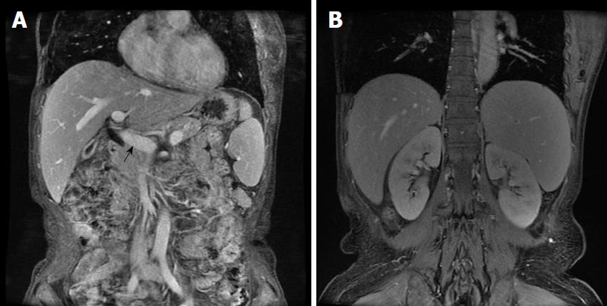
Figure 1 Abdominal changes shown by coronal magnetic resonance imaging.
A: Wider portal vein diameter (black arrow) shown in the coronal plane; B: Splenomegaly shown in the coronal plane.
- Citation: Yang QB, He YL, Peng CM, Qing YF, He Q, Zhou JG. Systemic lupus erythematosus complicated by noncirrhotic portal hypertension: A case report and review of literature. World J Clin Cases 2018; 6(13): 688-693
- URL: https://www.wjgnet.com/2307-8960/full/v6/i13/688.htm
- DOI: https://dx.doi.org/10.12998/wjcc.v6.i13.688


