Copyright
©The Author(s) 2017.
World J Clin Cases. Aug 16, 2017; 5(8): 340-343
Published online Aug 16, 2017. doi: 10.12998/wjcc.v5.i8.340
Published online Aug 16, 2017. doi: 10.12998/wjcc.v5.i8.340
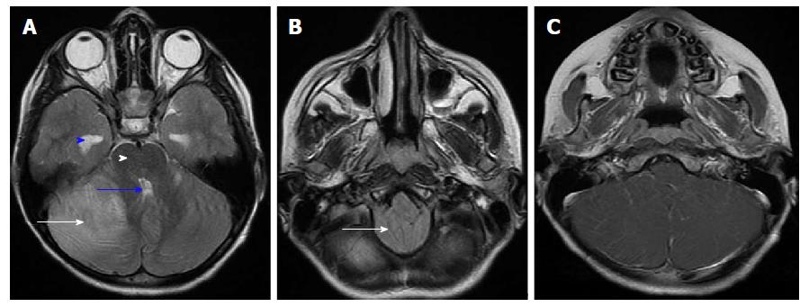
Figure 1 Magnetic resonance imaging: T2 and T1 post contrast images.
A and B: Cerebellar hyperintense areas on T2-weighted images predominant on the right side (white arrow in A), related to diffuse edema and producing cerebellar mass-effect on the fourth ventricle (blue arrow) and brainstem (white head arrow), tonsillar herniation (white arrow in B) and supratentorial hydrocephalus (blue head arrow); C: Gadolinium-enhanced T1-weighted sequence revealed leptomeningeal enhancement along the cerebellar folia.
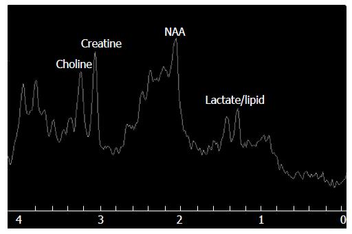
Figure 2 Magnetic resonance spectroscopy (TE = 35 ms) showed mildly reduced N actetyl aspartate/creatine and normal Choline/Creatine ratios.
Doublet of lactate/lipid peak (1.3 ppm) was detected. NAA: N actetyl aspartate.
- Citation: Ajmi H, Gaha M, Mabrouk S, Hassayoun S, Zouari N, Chemli J, Abroug S. Pseudotumoral acute cerebellitis associated with mumps infection in a child. World J Clin Cases 2017; 5(8): 340-343
- URL: https://www.wjgnet.com/2307-8960/full/v5/i8/340.htm
- DOI: https://dx.doi.org/10.12998/wjcc.v5.i8.340


