©The Author(s) 2016.
World J Clinical Cases. Sep 16, 2016; 4(9): 306-309
Published online Sep 16, 2016. doi: 10.12998/wjcc.v4.i9.306
Published online Sep 16, 2016. doi: 10.12998/wjcc.v4.i9.306
Figure 1 Computerized tomography scan revealing a large ovoid solid and cystic tumor (17 cm × 15 cm × 10 cm) at the left upper quadrant (right arrow).
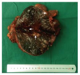
Figure 2 Macroscopically, a large ovoid solid and cystic tumor (20 cm × 20 cm × 10 cm) was observed at the left upper quadrant, and the tumor infiltrated the full-thickness wall of the jejunum (left arrow).
Figure 3 Macroscopically, an incision into the surface of the tumor revealed sclerotic tissue (up arrow) and bleeding regions inside the tumor (left arrow).
The tumor had infiltrated the full-thickness wall of the jejunum (right arrow).
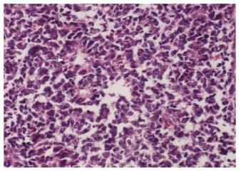
Figure 4 Small round cells containing uniform vesicular and Homer-Wright rosettes were found microscopically (right arrow, × 200).
Figure 5 Red blood cells were found between the tumor cells (right arrow, × 200).
- Citation: Liu Z, Xu YH, Ge CL, Long J, Du RX, Guo KJ. Huge peripheral primitive neuroectodermal tumor of the small bowel mesentery at nonage: A case report and review of the literature. World J Clinical Cases 2016; 4(9): 306-309
- URL: https://www.wjgnet.com/2307-8960/full/v4/i9/306.htm
- DOI: https://dx.doi.org/10.12998/wjcc.v4.i9.306


