©2014 Baishideng Publishing Group Inc.
World J Clin Cases. May 16, 2014; 2(5): 146-150
Published online May 16, 2014. doi: 10.12998/wjcc.v2.i5.146
Published online May 16, 2014. doi: 10.12998/wjcc.v2.i5.146
Figure 1 Ultrasound exam findings.
The images show the concentric thickening of the wall of the ascending colon, which assumed the appearance of a solid mass. In addition the big gallstone in the lumen of the gallbladder is displayed.
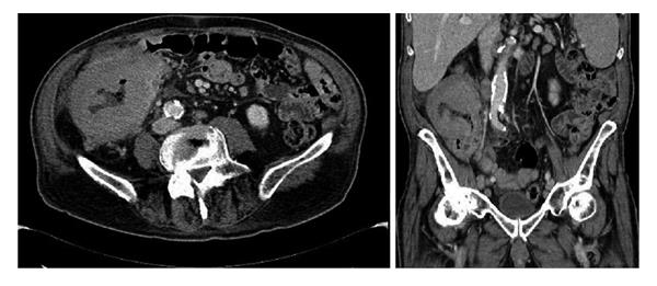
Figure 2 Computed tomography exam in the axial and coronal planes.
The images show the concentric thickening of the wall of the cecum and ascending colon. The millimetric air bubbles in the context of the colic wall are also depicted.
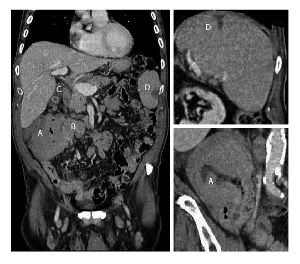
Figure 3 Coronal computed tomography scan (portal venous phase).
The images show the large colic lesion at the level of the hepatic flexure (A), the large necrotic lymphadenopathy close to the lesion (B), the stone in the lumen of the gallbladder (C) and the hypodense area in the context of the spleen indicative of the ischemic outcomes (D).
- Citation: Gigli S, Buonocore V, Barchetti F, Glorioso M, Di Brino M, Guerrisi P, Buonocore C, Giovagnorio F, Giraldi G. Primary colonic lymphoma: An incidental finding in a patient with a gallstone attack. World J Clin Cases 2014; 2(5): 146-150
- URL: https://www.wjgnet.com/2307-8960/full/v2/i5/146.htm
- DOI: https://dx.doi.org/10.12998/wjcc.v2.i5.146


