
















Copyright: ©Author(s) 2026.
World J Clin Cases. Oct 26, 2026; 14(28): 127546
Published online Oct 26, 2026. doi: 10.12998/wjcc.127546
Published online Oct 26, 2026. doi: 10.12998/wjcc.127546
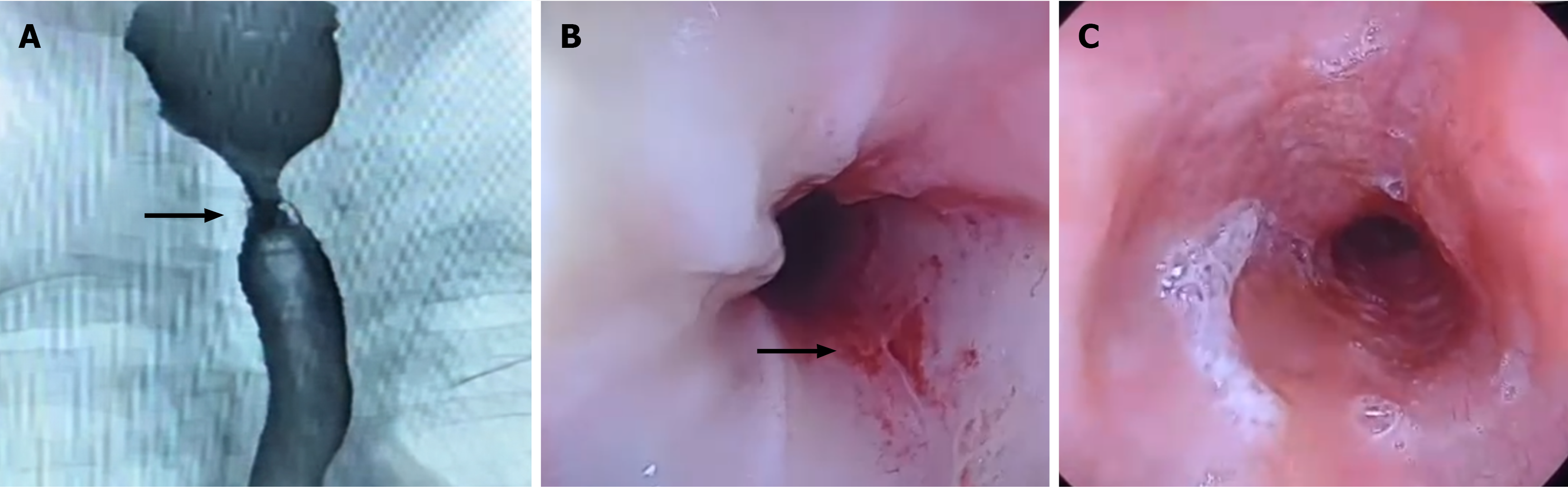
Figure 1 Radiological and endoscopic findings of esophageal stricture.
A: Barium swallow showing a short-segment stricture in the upper esophagus (black arrow); B: Upper gastrointestinal endoscopy showing luminal narrowing at 19 cm from the incisors, with mucosal erythema, fine nodularity, and ulceration (black arrow); C: Normal-appearing distal esophagus.
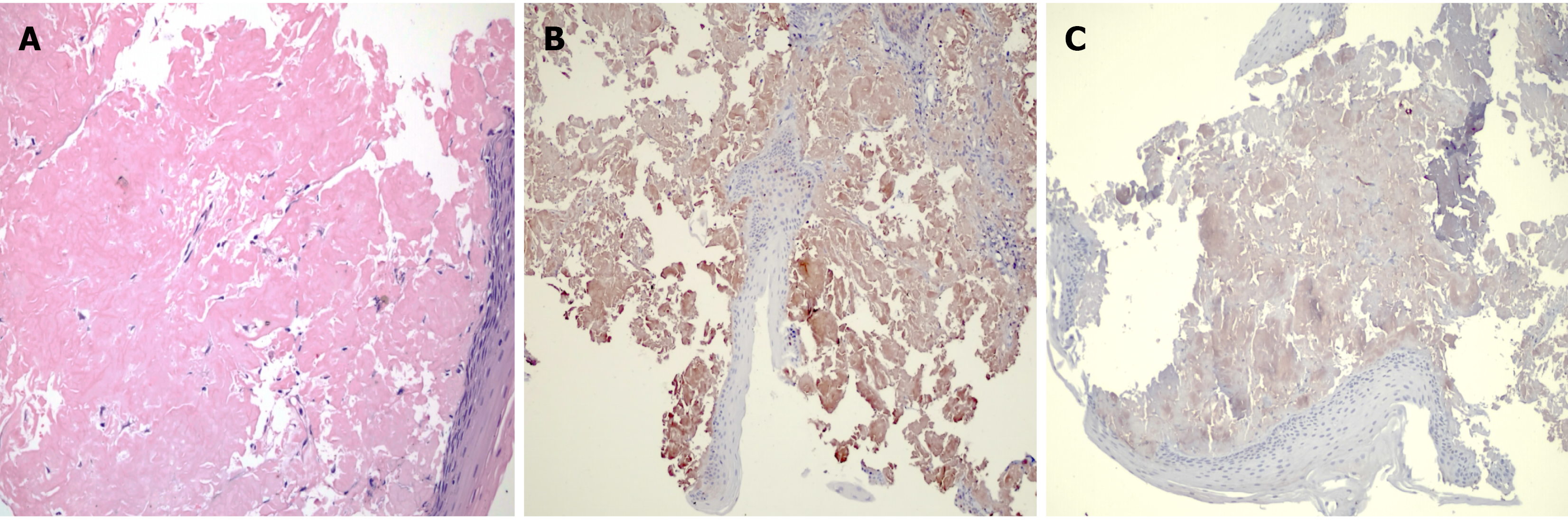
Figure 2 Histopathological and immunohistochemical findings of esophageal amyloid deposition.
A: Hematoxylin and eosin (H&E) staining showing abundant amorphous, eosinophilic extracellular material deposited within the subepithelial stroma, consistent with amyloid deposition (H&E ×100); B: Immunohistochemical staining showing positive cytoplasmic/extracellular staining for lambda light chains within the amyloid-containing subepithelial stroma (lambda ×40); C: Immunohistochemical staining showing negative kappa light-chain expression (Kappa ×40).
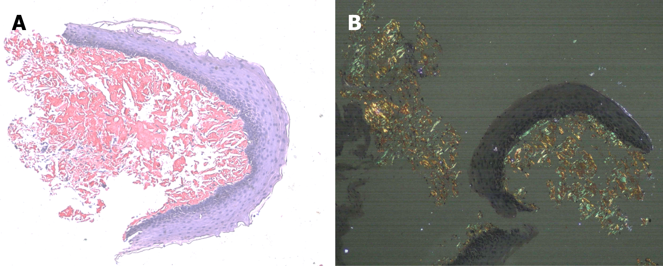
Figure 3 Congo red staining confirming amyloid deposition in the esophageal biopsy.
A: Congo red staining demonstrating salmon-pink amyloid deposits within the subepithelial stroma (×40); B: The same deposits showing characteristic apple-green birefringence under polarized light microscopy, confirming amyloid deposition (×40).
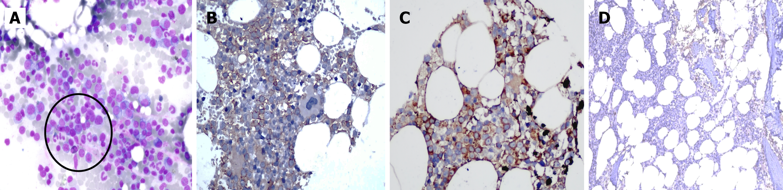
Figure 4 Bone marrow findings demonstrating an underlying plasma-cell dyscrasia.
A: Bone marrow aspirate smear showing increased mature and immature plasma cells with eccentrically placed nuclei, abundant basophilic cytoplasm, and perinuclear hof (black circle); residual erythroid and myeloid precursors are also present (Giemsa ×200); B: Immunohistochemical staining shows plasma cells with membranous CD138 expression (x100); C: The neoplastic plasma cells demonstrated positive expression for lambda light-chain (×100); D: Negative for kappa light chain, indicating lambda light chain restriction (×100) .
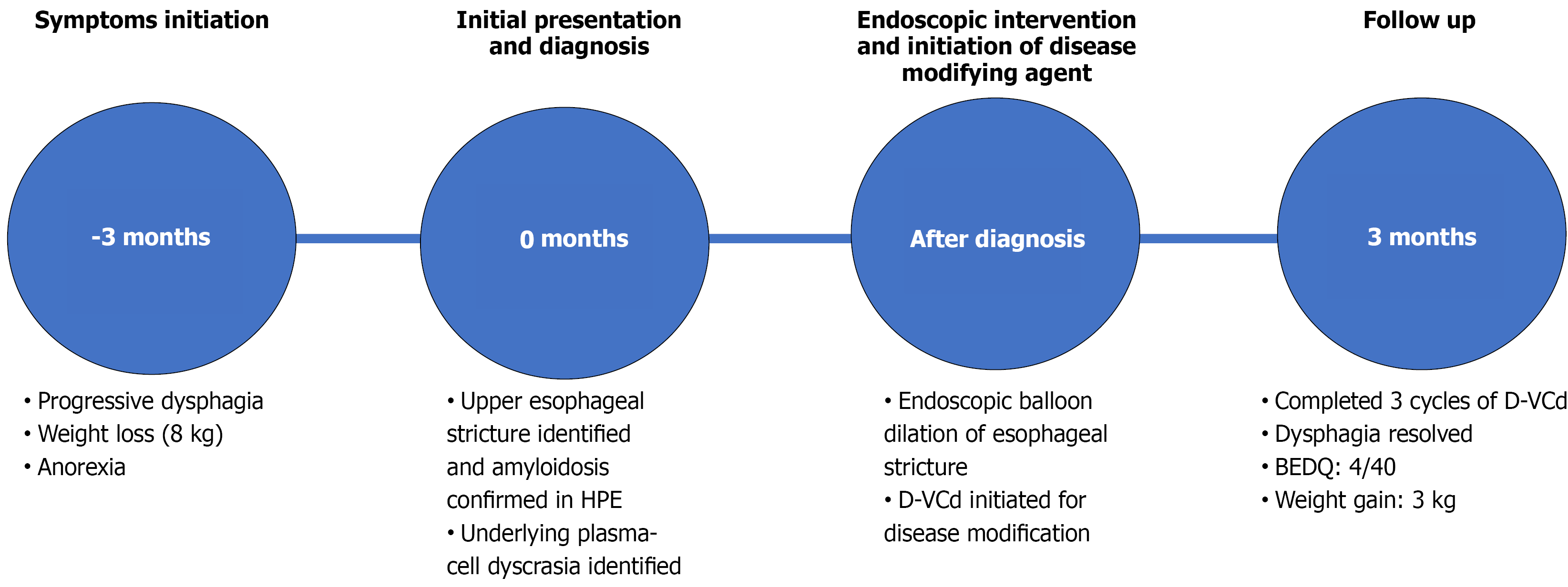
Figure 5 Timeline of clinical events and management.
Timeline depicting the major clinical, diagnostic, therapeutic, and follow-up events during the patient’s clinical course. D-VCd: Daratumumab, bortezomib, cyclophosphamide, and dexamethasone; BEDQ: Brief Esophageal Dysphagia Questionnaire; HPE: Histopathological examination.
- Citation: Sachdeva S, Sirohi N, Mandal K, Bansal D, Vaithiyam V, Dalal A, Goyal S, Sakhuja P. Esophageal stricture due to amyloidosis in plasma cell dyscrasia: A case report and review of literature. World J Clin Cases 2026; 14(28): 127546
- URL: https://www.wjgnet.com/2307-8960/full/v14/i28/127546.htm
- DOI: https://dx.doi.org/10.12998/wjcc.127546