
















Copyright: ©Author(s) 2026.
World J Clin Cases. Oct 26, 2026; 14(28): 125577
Published online Oct 26, 2026. doi: 10.12998/wjcc.125577
Published online Oct 26, 2026. doi: 10.12998/wjcc.125577

Figure 1 Dental/periodontal clinical examination.
A: Frontal intraoral view demonstrating the three-unit ceramometal fixed dental prosthesis (FDP) in situ and the associated marginal inflammation; B: Palatal close-up view showing the defective prosthetic margins and the exophytic lesion emerging from the gingival crevice; C: Occlusal view following removal of the FDP, revealing the exophytic palatal lesion originating beneath the ill-fitting prosthesis.
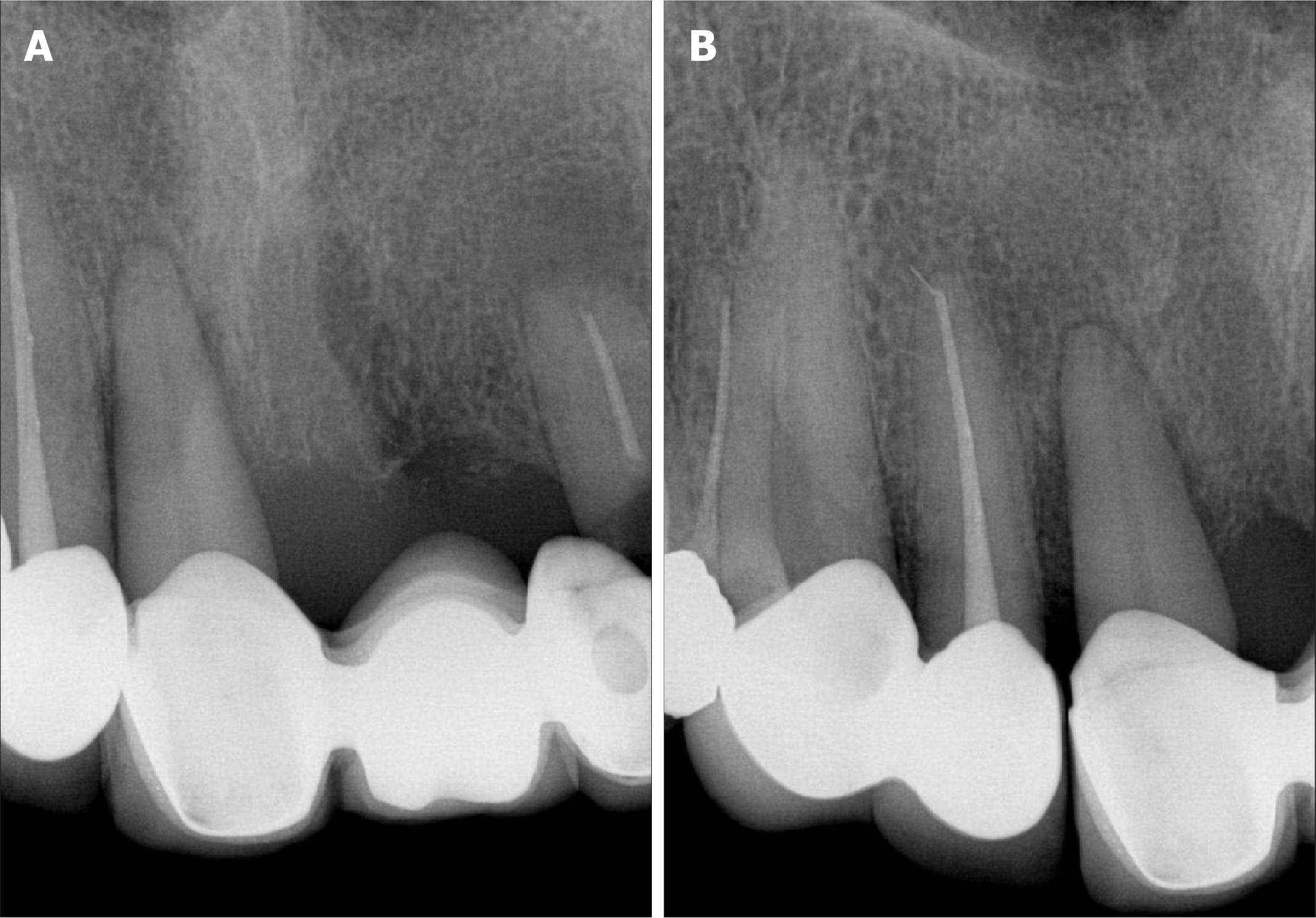
Figure 2 Imaging examination.
A: Periapical radiograph of the right maxillary anterior region demonstrating severe alveolar bone loss; B: Periapical radiograph of the left maxillary anterior region showing the pattern of bone loss and widening of the periodontal ligament space.
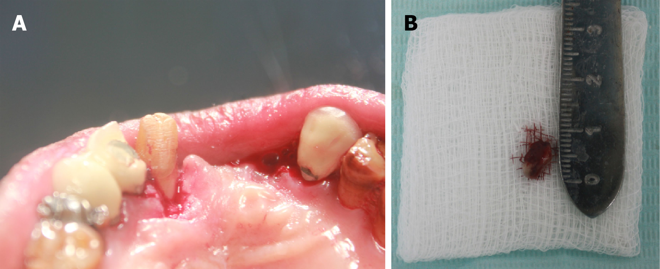
Figure 3 Intraoperative view and gross specimen photograph.
A: Intraoperative view of the surgical site immediately following complete excision of the lesion and extraction of tooth #22; B: Gross specimen photograph of the excised lesion placed adjacent to a ruler, measuring approximately 1.0 cm.
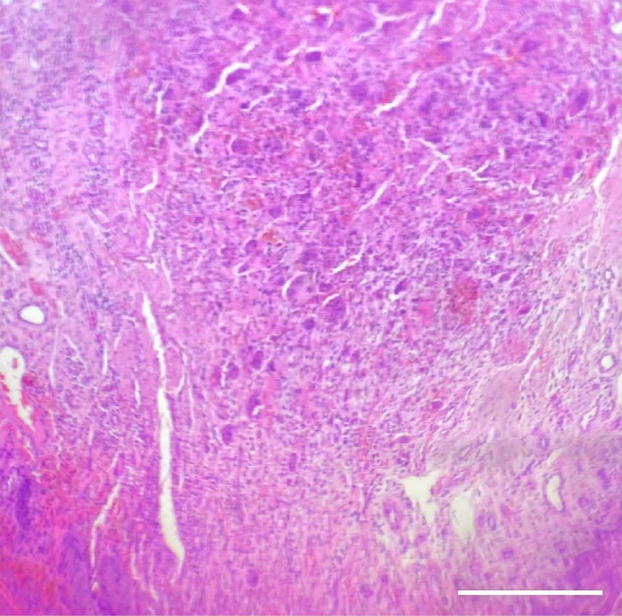
Figure 4 Hematoxylin and eosin stained photomicrograph showing numerous multinucleated giant cells within a cellular vascular stroma (original magnification ×200).
Scale bar = 100 µm.
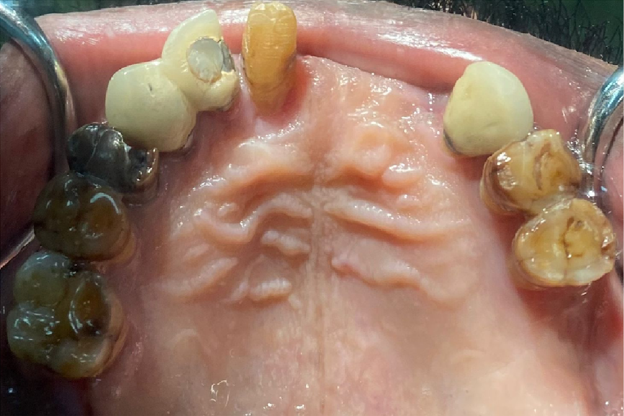
Figure 5 Occlusal view of the maxillary arch at the 20-day postoperative follow-up appointment demonstrating complete mucosal healing.
- Citation: Aljabr AA, Dobaian KS. Peripheral giant cell granuloma associated with dental prostheses: A case report. World J Clin Cases 2026; 14(28): 125577
- URL: https://www.wjgnet.com/2307-8960/full/v14/i28/125577.htm
- DOI: https://dx.doi.org/10.12998/wjcc.125577