
















Copyright: ©Author(s) 2026.
World J Clin Cases. Sep 6, 2026; 14(25): 124208
Published online Sep 6, 2026. doi: 10.12998/wjcc.124208
Published online Sep 6, 2026. doi: 10.12998/wjcc.124208
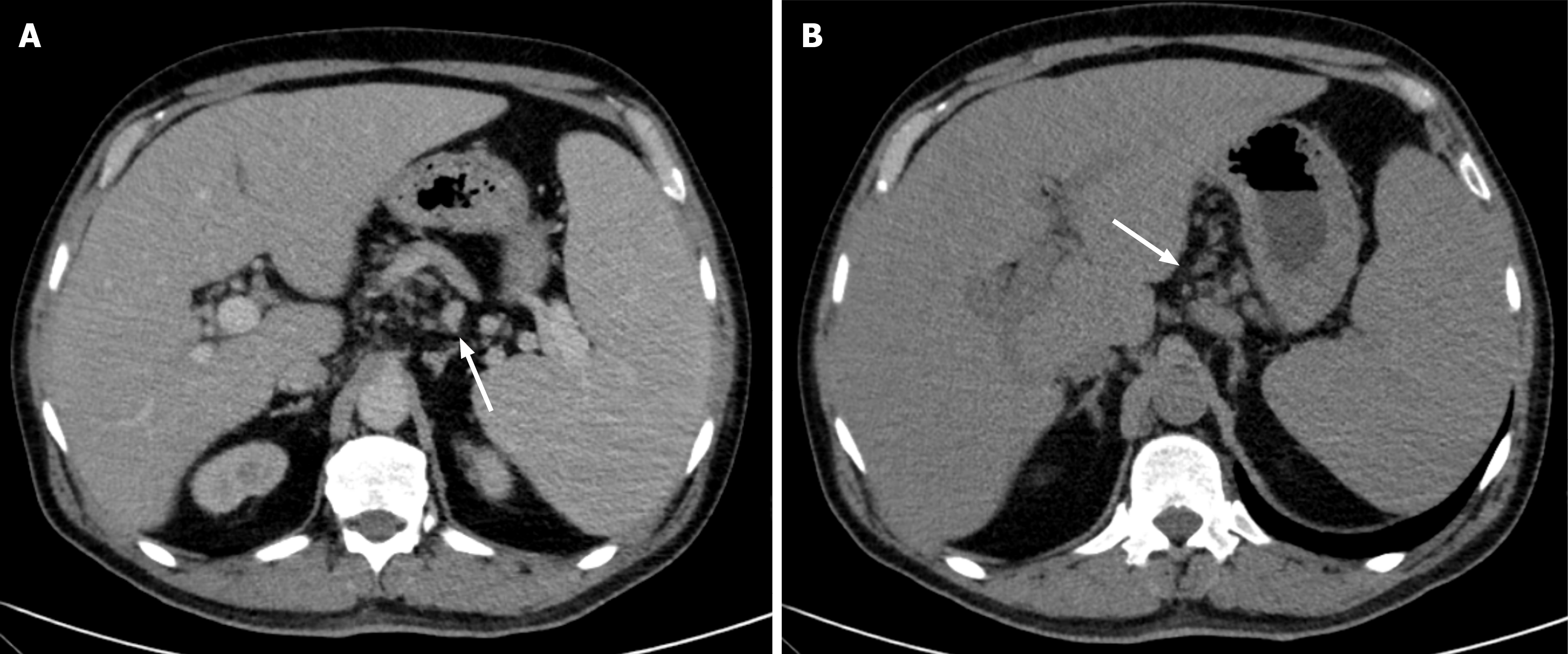
Figure 1 Contrast-enhanced computed tomography image.
A: The abdomen shows enlarged liver and spleen, and a cluster of lymph nodes (white arrow); B: The abdomen shows mesenteric lymphadenopathy (white arrow).
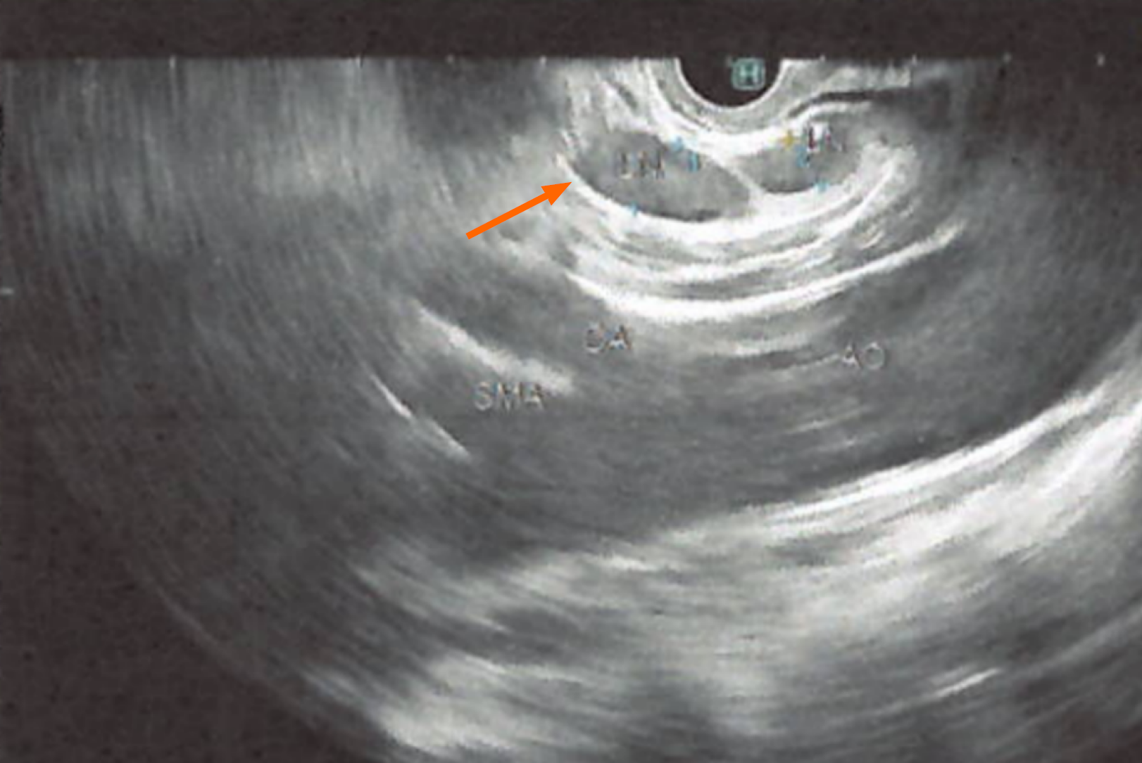
Figure 2 Endoscopic ultrasound image demonstrates left gastric territory lymph nodes (orange arrow).
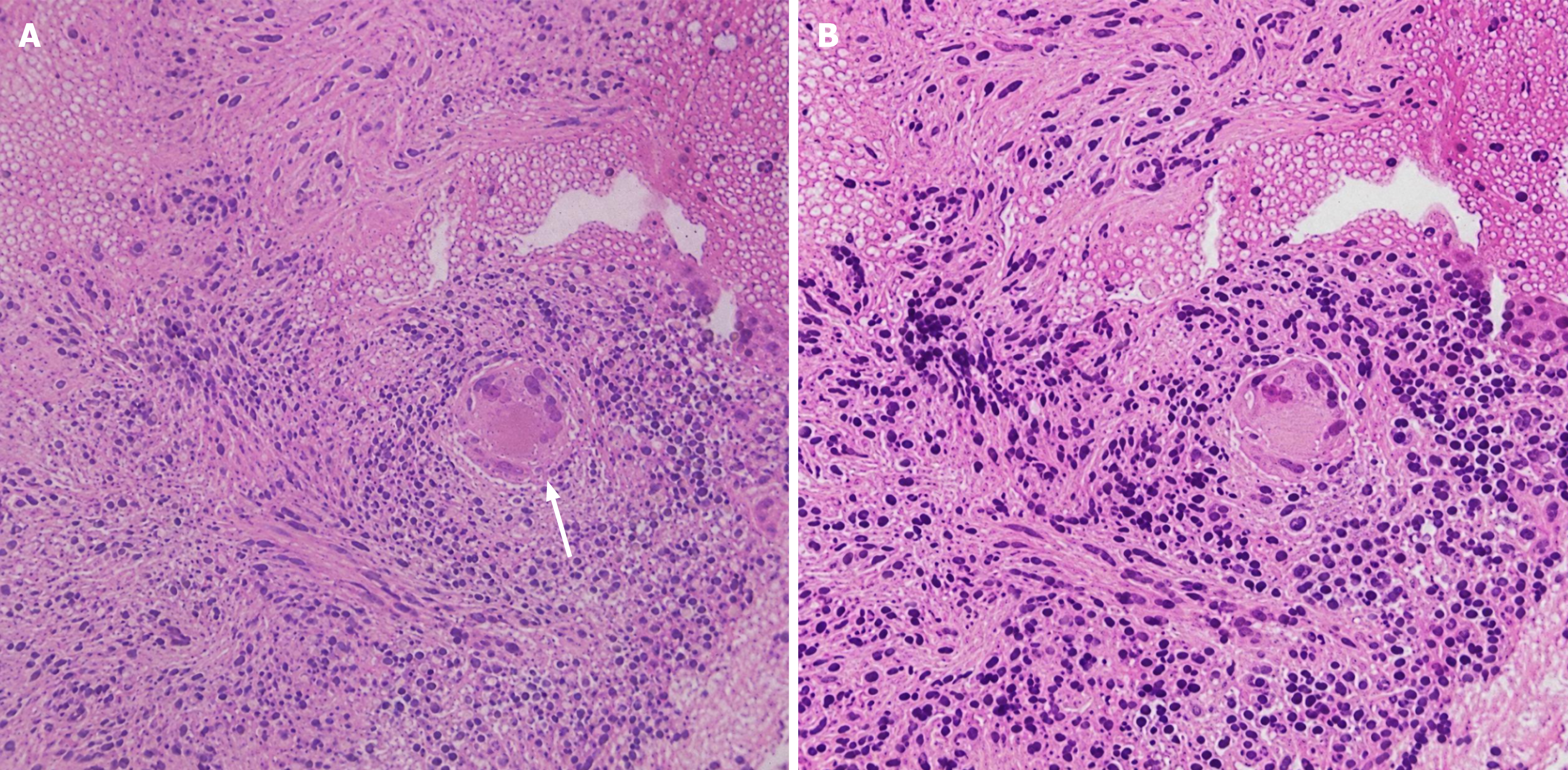
Figure 3 Histopathological image demonstrates noncaseating granulomatous inflammation with multinucleated giant cells, consistent with sarcoidosis.
A: White arrow pointing at noncaseating granuloma; B: Magnified picture of histopathological slide.
- Citation: Almasaabi M, Masood S, Elsayed G. Concurrent sarcoidosis and hemophagocytic lymphohistiocytosis: A case report. World J Clin Cases 2026; 14(25): 124208
- URL: https://www.wjgnet.com/2307-8960/full/v14/i25/124208.htm
- DOI: https://dx.doi.org/10.12998/wjcc.124208