











Copyright: ©Author(s) 2026.
World J Clin Cases. Aug 26, 2026; 14(24): 122937
Published online Aug 26, 2026. doi: 10.12998/wjcc.122937
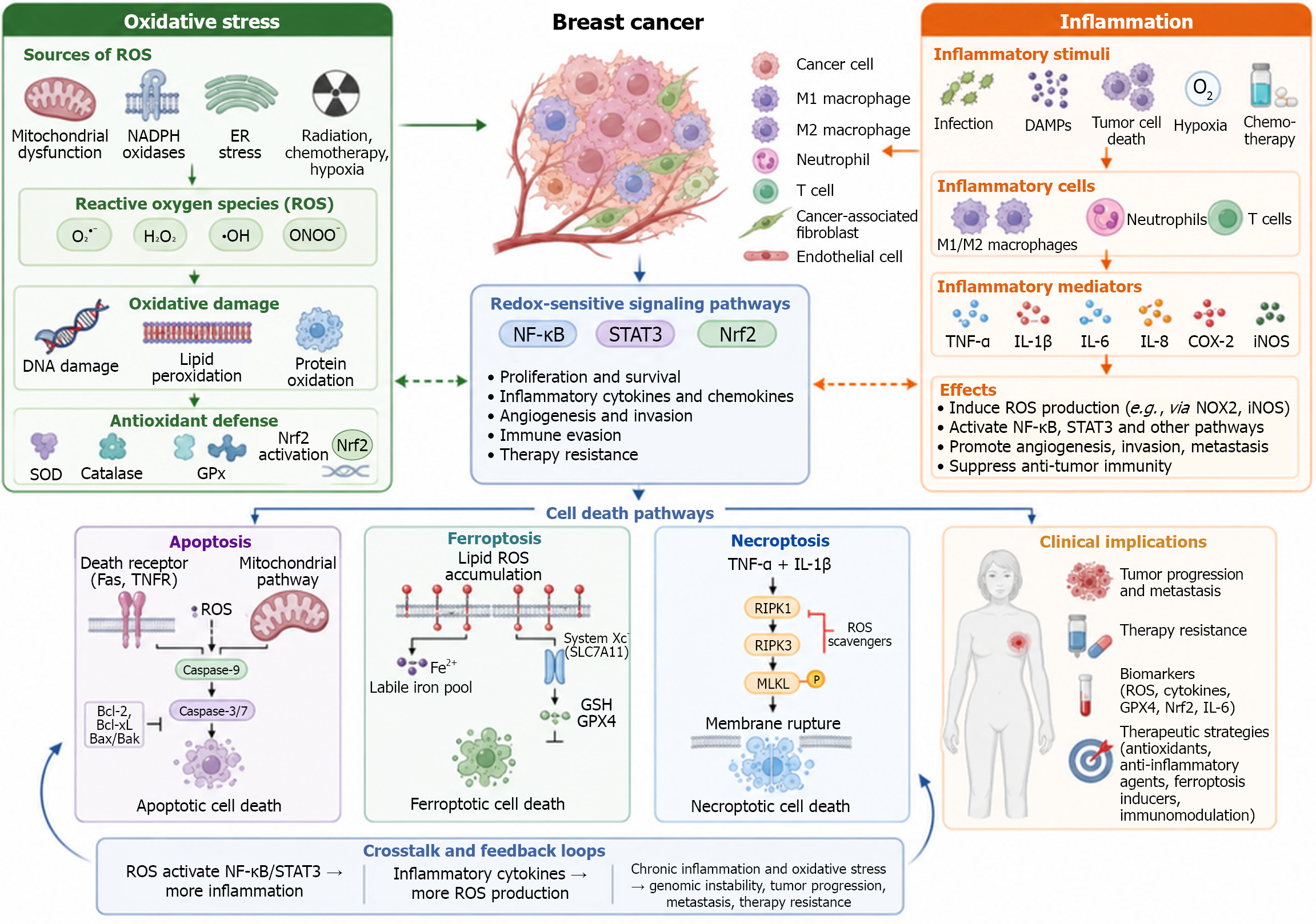
Figure 1 Interplay among oxidative stress, inflammation, and regulated cell death pathways in breast cancer.
The left panel illustrates major sources of reactive oxygen species (ROS), including mitochondrial dysfunction, NADPH oxidase 2 (NOX2) activation, endoplasmic reticulum stress, radiation, chemotherapy, and hypoxia. Accumulation of ROS, such as superoxide, hydrogen peroxide, hydroxyl radicals, and peroxynitrite, promotes DNA damage, lipid peroxidation, and protein oxidation. These effects are counteracted by antioxidant defense systems, including superoxide dismutase, catalase, glutathione (GSH) peroxidase, and nuclear factor erythroid 2-related factor 2 (Nrf2)-mediated transcriptional responses. The right panel summarizes inflammatory stimuli, including infection, damage-associated molecular patterns, tumor cell death, hypoxia, and anticancer therapy, which activate macrophages, neutrophils, and T cells. These immune cells release inflammatory mediators such as tumor necrosis factor-α, interleukin (IL)-1β, IL-6, IL-8, cyclooxygenase-2, and inducible nitric oxide synthase (iNOS), thereby enhancing ROS generation, angiogenesis, invasion, metastasis, and suppression of antitumor immunity. The central panel shows the micro environment of breast cancer (BC), composed of tumor cells, M1 and M2 macrophages, neutrophils, T cells, cancer-associated fibroblasts, and endothelial cells, highlighting their dynamic interactions with oxidative stress and inflammatory signaling. In the BC microenvironment, oxidative and inflammatory signals converge on redox-sensitive pathways, particularly nuclear factor kappa B (NF-κB), signal transducer and activator of transcription 3 (STAT3), and Nrf2, which regulate tumor-cell proliferation, survival, immune evasion, angiogenesis, and therapeutic resistance. ROS can activate NF-κB and STAT3, increasing the production of inflammatory cytokines, whereas cytokines and inflammatory enzymes stimulate additional ROS production through pathways involving NOX2 and iNOS. This reciprocal interaction establishes a self-amplifying feedback loop that sustains chronic inflammation and oxidative stress. Depending on the intensity, duration, and cellular context of these signals, different regulated cell death pathways may be activated. Apoptosis is mediated through death-receptor and mitochondrial pathways involving caspase-9, caspase-3/7, and Bcl-2 family proteins. Ferroptosis results from iron-dependent lipid ROS accumulation and is regulated by the system Xc-GSH-GPX4 antioxidant axis. Necroptosis is mediated by RIPK1, RIPK3, and phosphorylated MLKL, leading to membrane rupture and the release of inflammatory intracellular contents. Although activation of these pathways may eliminate tumor cells, cancer cells can adapt by increasing antioxidant defenses, altering death-signaling proteins, and activating survival pathways, thereby promoting treatment resistance. Collectively, the crosstalk among oxidative stress, inflammation, and cell death contributes to genomic instability, tumor progression, metastasis, biomarker alterations, and variable therapeutic responses, while also providing potential targets for antioxidant, anti-inflammatory, ferroptosis-inducing, and immunomodulatory strategies. ROS: Reactive oxygen species; NADPH: Nicotinamide adenine dinucleotide phosphate; ER: Endoplasmic reticulum; SOD: Superoxide dismutase; GPx: Glutathione peroxidase; Nrf2: Nuclear factor erythroid 2-related factor 2; M1: Classically activated pro-inflammatory macrophage phenotype; M2: Alternatively activated anti-inflammatory macrophage phenotype; DAMPs: Damage-associated molecular patterns; TNF-α: Tumor necrosis factor-α; IL: Interleukin; COX-2: Cyclooxygenase-2; iNOS: Inducible nitric oxide synthase; NOX2: NADPH oxidase 2; NF-κB: Nuclear factor kappa B; STAT3: Signal transducer and activator of transcription 3; Fas: Fas cell surface death receptor; TNFR: Tumor necrosis factor receptor; Bcl-2: B-cell lymphoma 2; Bcl-xL: B-cell lymphoma-extra large; Bax: BCL2-associated X protein; Bak: BCL2 antagonist/killer 1; Fe2+: Ferrous iron; System Xc-: Cystine/glutamate antiporter system Xc-; SLC7A11: Solute carrier family 7 member 11; GSH: Glutathione; GPX4: Glutathione peroxidase 4; RIPK1: Receptor-interacting serine/threonine-protein kinase 1; RIPK3: Receptor-interacting serine/threonine-protein kinase 3; MLKL: Mixed lineage kinase domain-like pseudokinase. This figure was created by BioRender.com (Supplementary material).
- Citation: Tutar Ş, Uçar Çifçi K, Tutar Y. Oxidative stress and inflammation in breast cancer: Mechanistic interactions and clinical implications. World J Clin Cases 2026; 14(24): 122937
- URL: https://www.wjgnet.com/2307-8960/full/v14/i24/122937.htm
- DOI: https://dx.doi.org/10.12998/wjcc.122937