
















Copyright: ©Author(s) 2026.
World J Clin Cases. Jul 16, 2026; 14(20): 121605
Published online Jul 16, 2026. doi: 10.12998/wjcc.121605
Published online Jul 16, 2026. doi: 10.12998/wjcc.121605
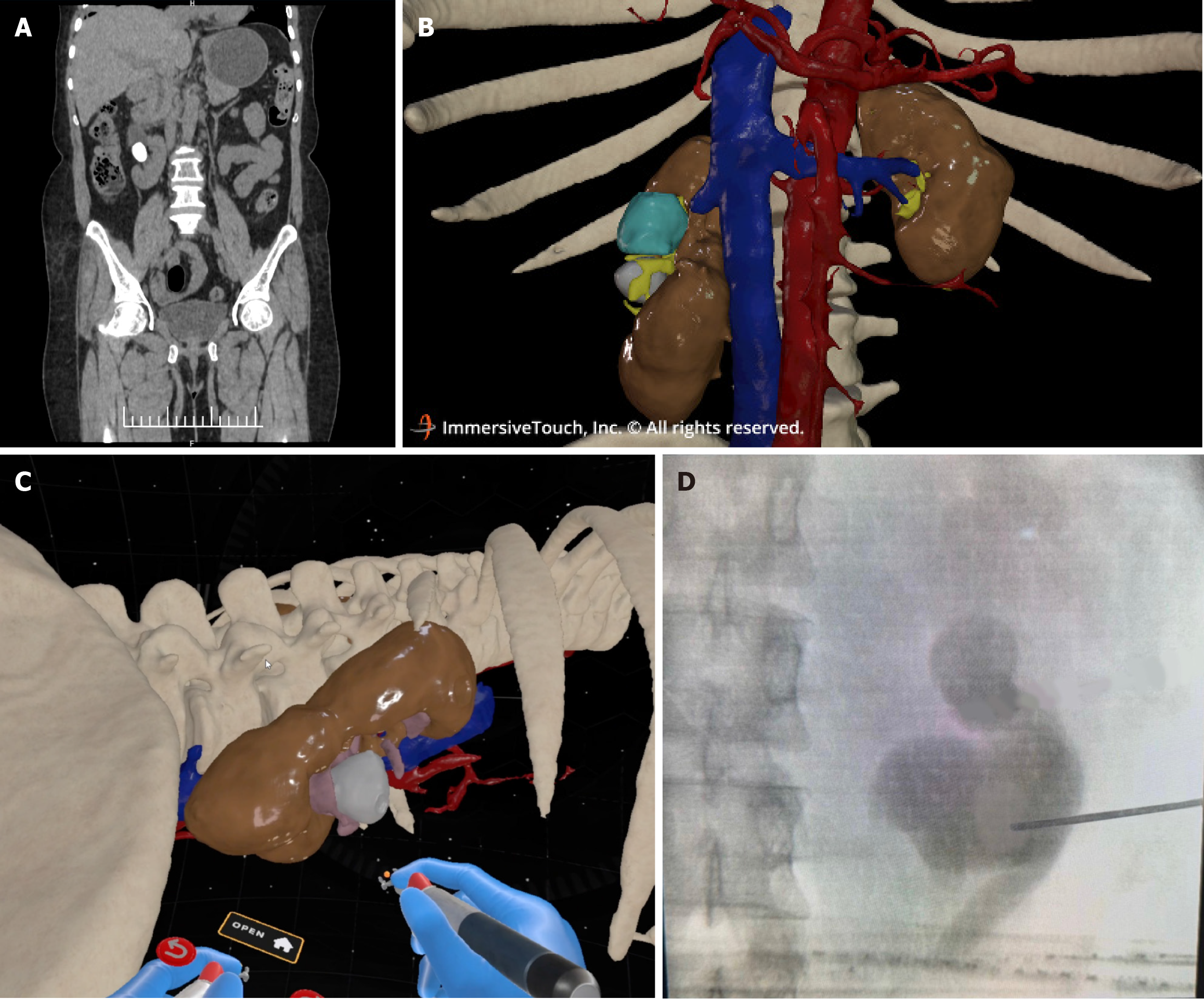
Figure 1 Multimodal imaging and virtual reality–guided planning of percutaneous nephrolithotomy in a malrotated kidney.
A: Contrast-enhanced computed tomography coronal image demonstrating a malrotated right kidney with an anterolaterally oriented renal hilum and a hyperdense renal pelvic calculus; B: Three-dimensional reconstruction generated from computed tomography DICOM data illustrating patient-specific renal anatomy, including the malrotated kidney, renal vasculature, and surrounding skeletal landmarks; C: Virtual reality–based immersive visualization used for preoperative planning, enabling identification of the posterior lower calyx and simulation of the planned posterior lower calyceal subcostal access tract; D: Intraoperative fluoroscopic image confirming successful posterior lower calyceal puncture corresponding to the preoperatively planned virtual reality trajectory.
- Citation: Kothapalli S, Sriskandaraja T, Palaniyandi V, Sekar H, Krishnamoorthy S. Virtual reality-enabled precision in percutaneous nephrolithotomy - strategic access planning in a malrotated kidney: A case report. World J Clin Cases 2026; 14(20): 121605
- URL: https://www.wjgnet.com/2307-8960/full/v14/i20/121605.htm
- DOI: https://dx.doi.org/10.12998/wjcc.121605