
















Copyright: ©Author(s) 2026.
World J Clin Cases. Jun 6, 2026; 14(16): 120049
Published online Jun 6, 2026. doi: 10.12998/wjcc.v14.i16.120049
Published online Jun 6, 2026. doi: 10.12998/wjcc.v14.i16.120049
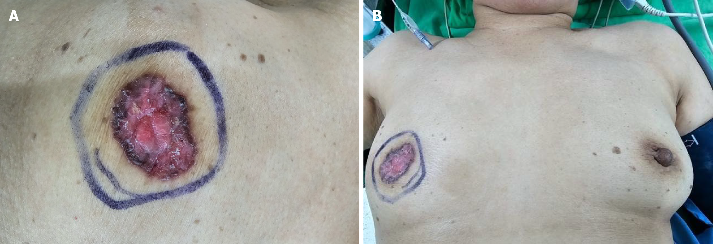
Figure 1 Clinical presentation of an ulcerated right nipple lesion.
A: Close-up view of an ulcerative lesion of the right nipple characterized by erythema, crusting, and irregular margins, confined to the outlined area; B: Wider field view showing involvement of the right nipple-areolar complex without extension into the surrounding breast parenchyma or the contralateral breast. The left breast appeared normal, with no similar lesions.
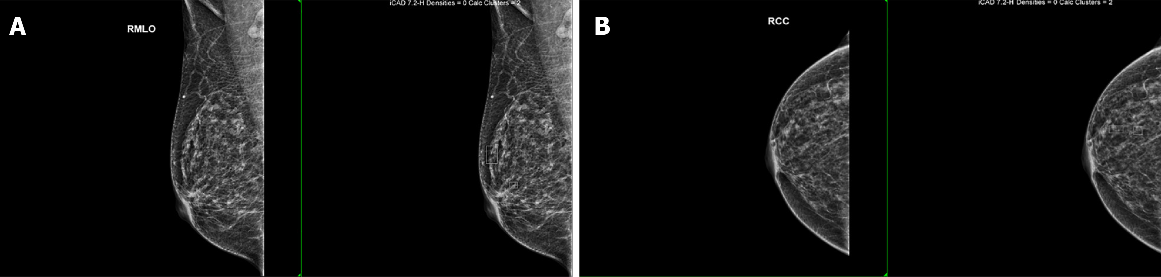
Figure 2 Breast mammography.
A: Right breast mammography. Right craniocaudal and right mediolateral oblique views demonstrated grouped punctate and amorphous calcifications in the upper outer quadrant and retroareolar region of the right breast, without associated mass, architectural distortion, or lym
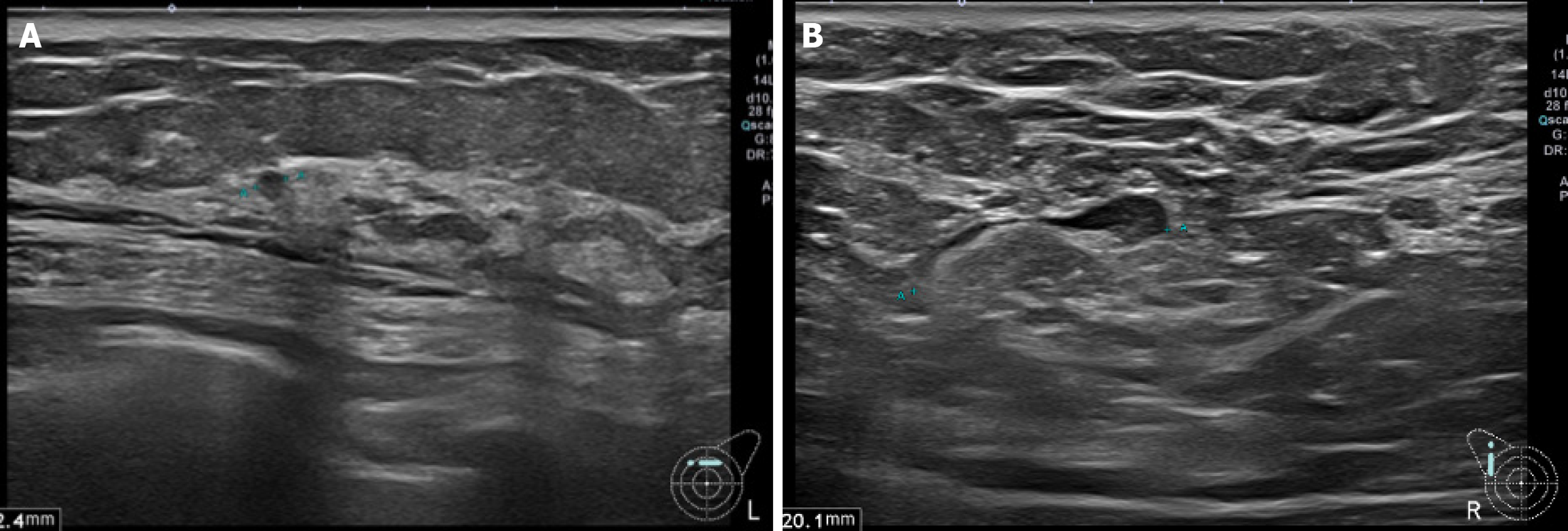
Figure 3 Bilateral breast ultrasonography.
Ultrasonography demonstrated scattered subcentimeter hypoechoic nodules (< 5 mm) in both breasts, most consistent with benign complex cysts or fibrocystic changes.
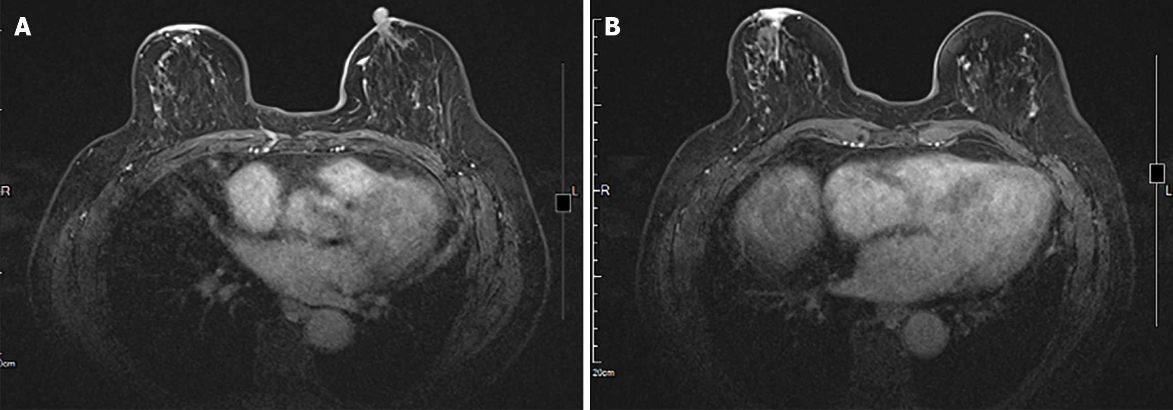
Figure 4 Magnetic resonance imaging.
A: Magnetic resonance imaging (MRI) of the right nipple-areolar complex. Axial contrast-enhanced MRI demonstrated right nipple inversion with associated skin thickening and enhancement, consistent with Paget’s disease; B: Bilateral breast MRI. Multiple small enhancing foci are present in both breasts, consistent with benign fibrocystic changes.
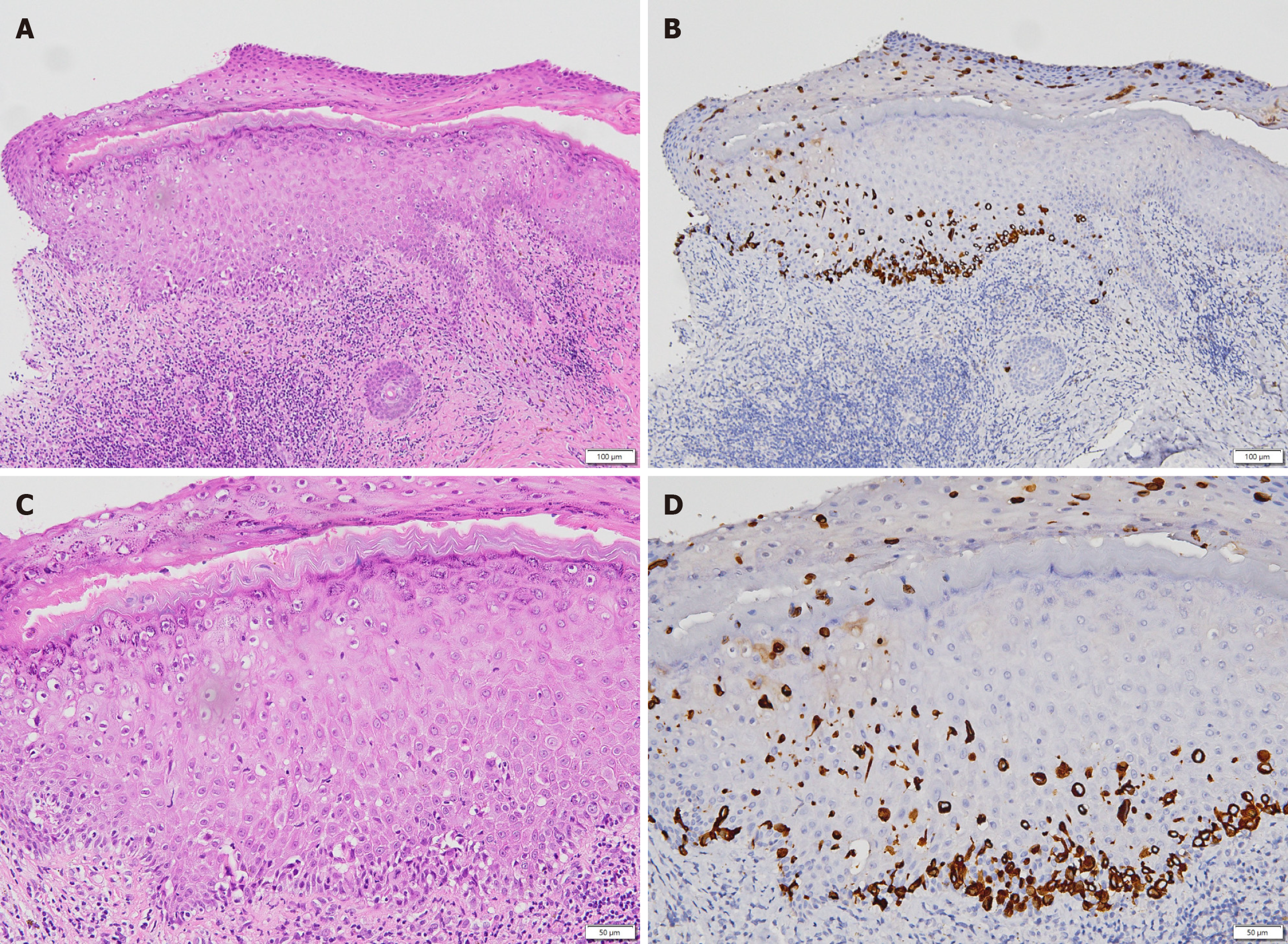
Figure 5 Histopathologic features of Paget’s disease of the nipple.
A: Skin, 40 µm; B: Cytokeratin 7 (CK7) immunostaining, 40 µm; C: Skin, 100 µm; D: CK7 immunostaining, 100 µm. Microscopic examination demonstrated malignant ductal epithelial cells spreading along the basal layer and infiltrating upward into the overlying squamous epithelium. Paget cells were ovoid, with high nuclear-to-cytoplasmic ratios, hyperchromatic nuclei, and pale cytoplasm. Immunohistochemistry showed strong CK7 positivity, supporting the diagnosis.
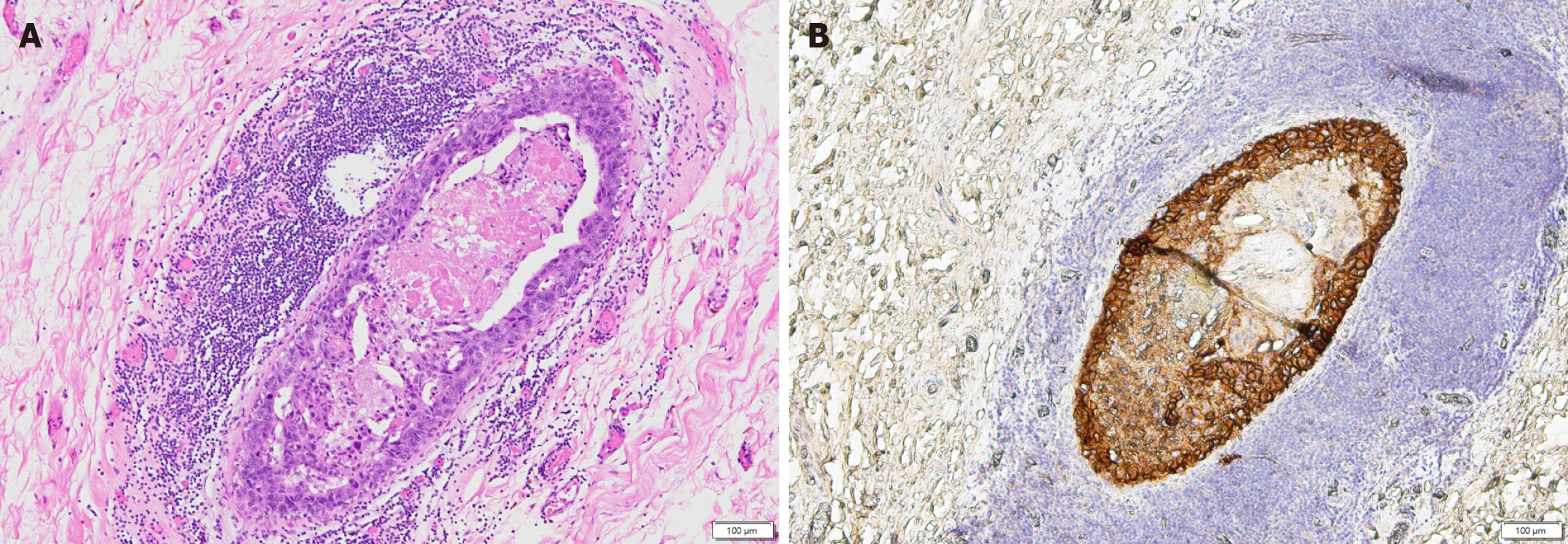
Figure 6 High-grade ductal carcinoma in situ in the partial mastectomy specimen.
A and B: Representative sections are shown which demonstrated monotonous tumor cells with enlarged, hyperchromatic nuclei filling and expanding mammary ducts. Central comedo-type necrosis and peripheral reactive lymphoid aggregates were present, consistent with high-grade ductal carcinoma in situ.
- Citation: Tsai CY, Lo TC, Yu JC, Hong ZJ, Sung YK. Paget’s disease of the nipple with occult ductal carcinoma in situ despite negative conventional imaging: A case report. World J Clin Cases 2026; 14(16): 120049
- URL: https://www.wjgnet.com/2307-8960/full/v14/i16/120049.htm
- DOI: https://dx.doi.org/10.12998/wjcc.v14.i16.120049