©The Author(s) 2025.
World J Clin Cases. Nov 6, 2025; 13(31): 109584
Published online Nov 6, 2025. doi: 10.12998/wjcc.v13.i31.109584
Published online Nov 6, 2025. doi: 10.12998/wjcc.v13.i31.109584
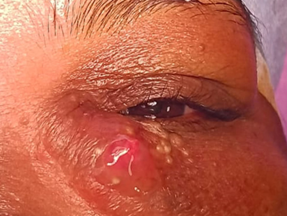
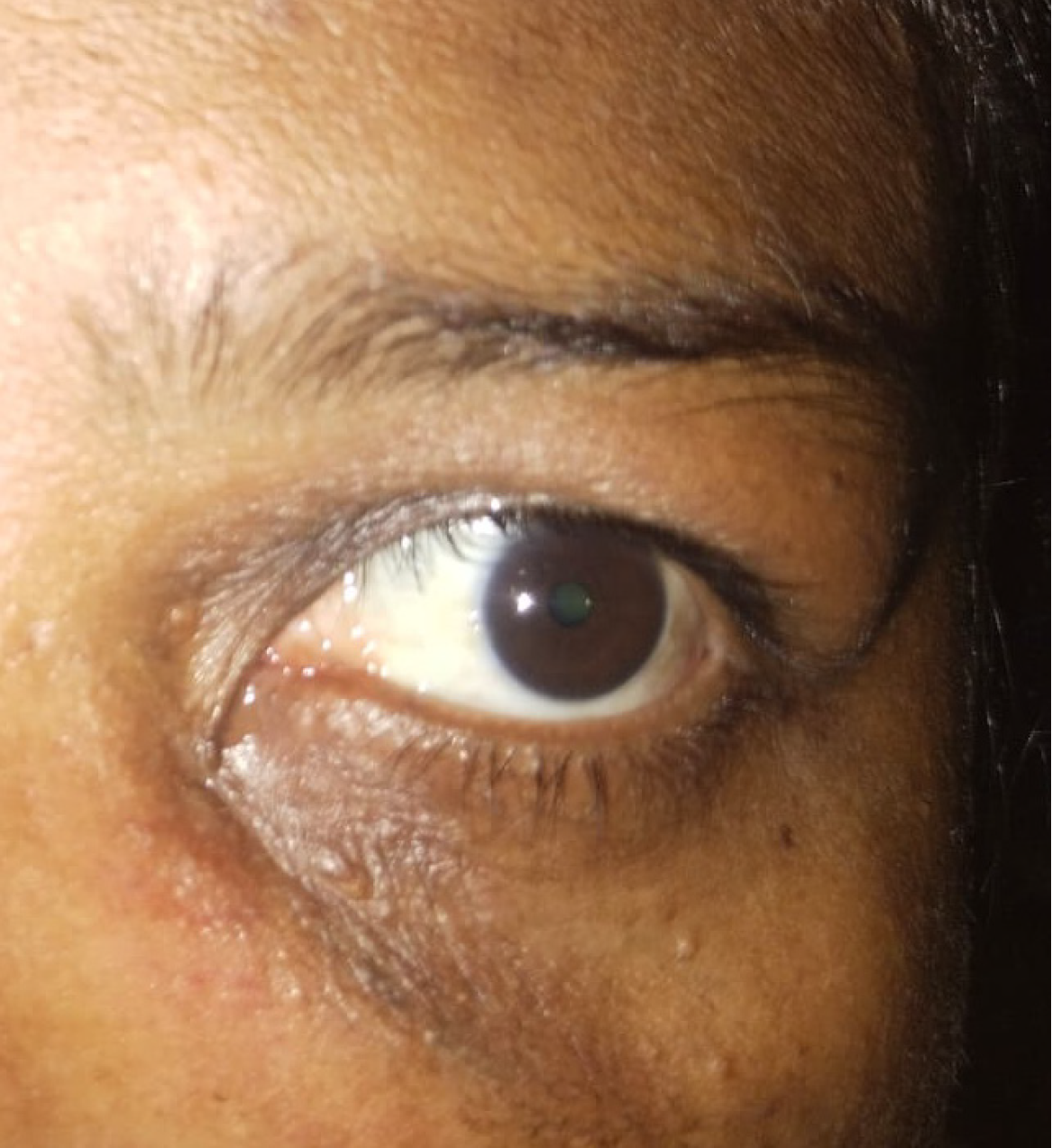
Figure 1 Clinical photo.
The photo shows swelling over the left medial canthal area with overlying skin showing puckered appearance, white comedones, and a fistulous opening with mucoid discharge.
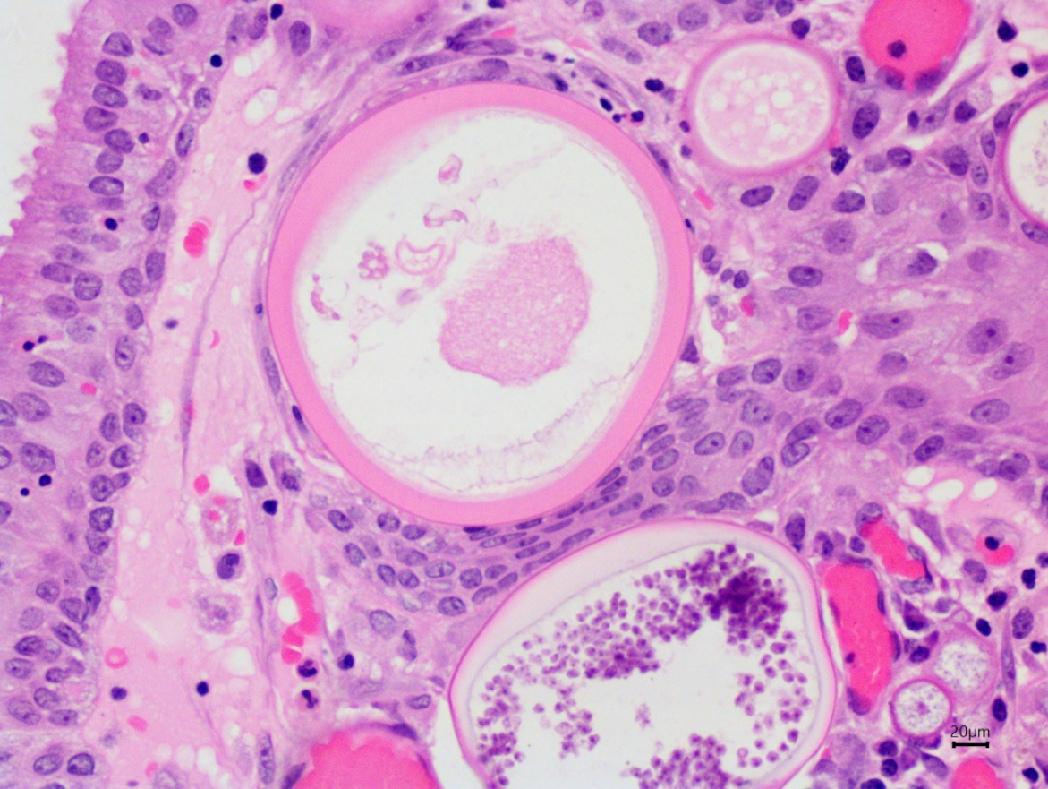
Figure 2 Histopathological examination.
The excised mass showed thick-walled sporangia rupturing to release endospores, suggestive of rhinosporidiosis.
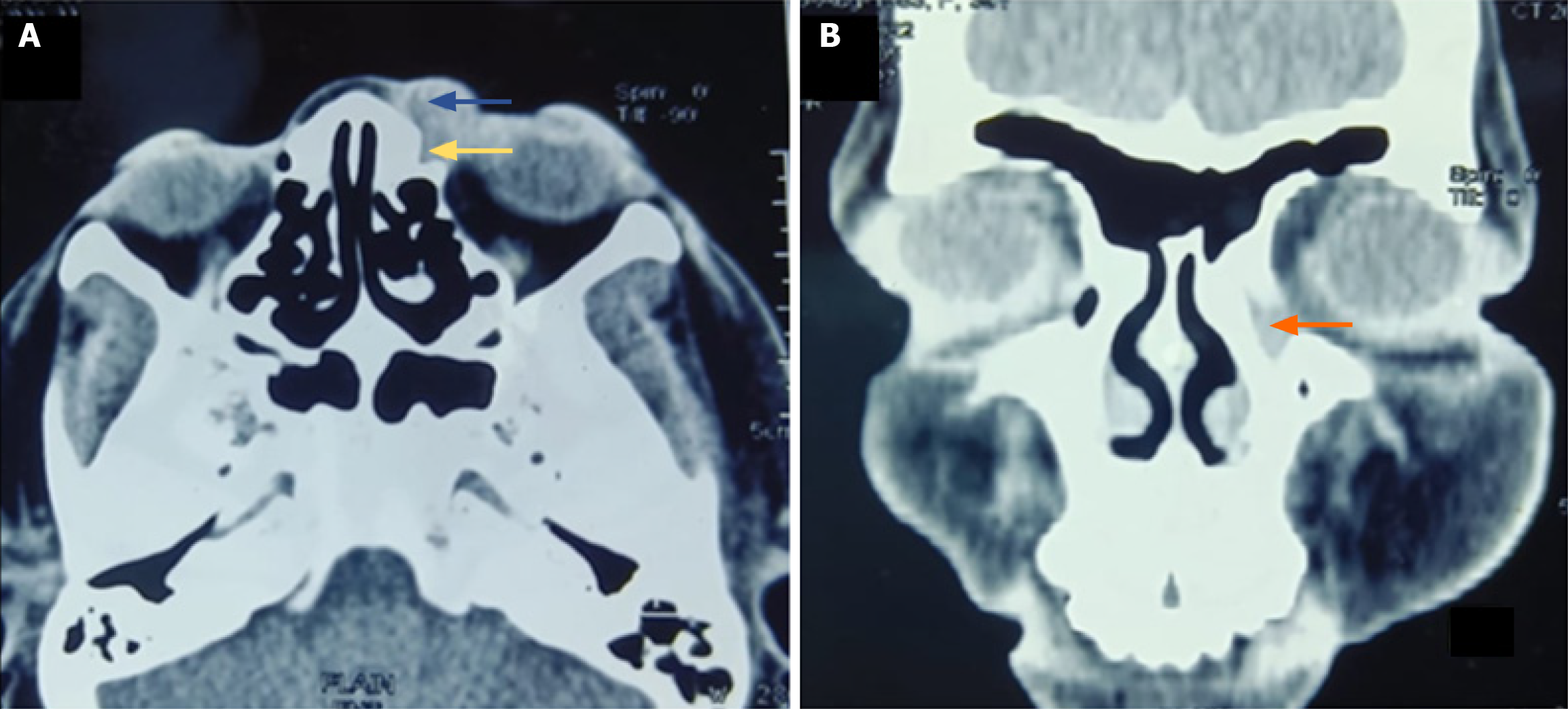
Figure 3 Computed tomography.
A and B: Isodense mass lesion in lacrimal sac area showing extension into preseptal area (marked with yellow arrow), left nasolacrimal duct (marked with orange arrow).
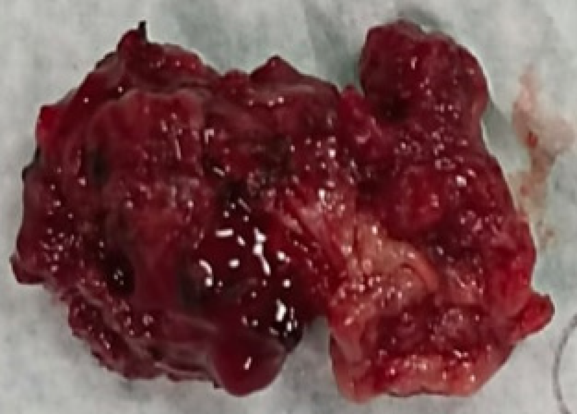
Figure 4 Gross specimen of the surgically excised mass.
The show a strawberry-like polypoidal vascular mass, with multiple papillary excrescences.
Figure 5 Clinical photo.
The photo shows a healthy periocular area near medial canthus, with the patient showing no recurrence at 3 years postoperatively.
- Citation: Panda BB, Kar A, Koppalu Lingaraju T, Ayyanar P. Acquired cutaneous fistula in the periocular area: A case report. World J Clin Cases 2025; 13(31): 109584
- URL: https://www.wjgnet.com/2307-8960/full/v13/i31/109584.htm
- DOI: https://dx.doi.org/10.12998/wjcc.v13.i31.109584


