©The Author(s) 2025.
World J Clin Cases. Oct 26, 2025; 13(30): 112419
Published online Oct 26, 2025. doi: 10.12998/wjcc.v13.i30.112419
Published online Oct 26, 2025. doi: 10.12998/wjcc.v13.i30.112419
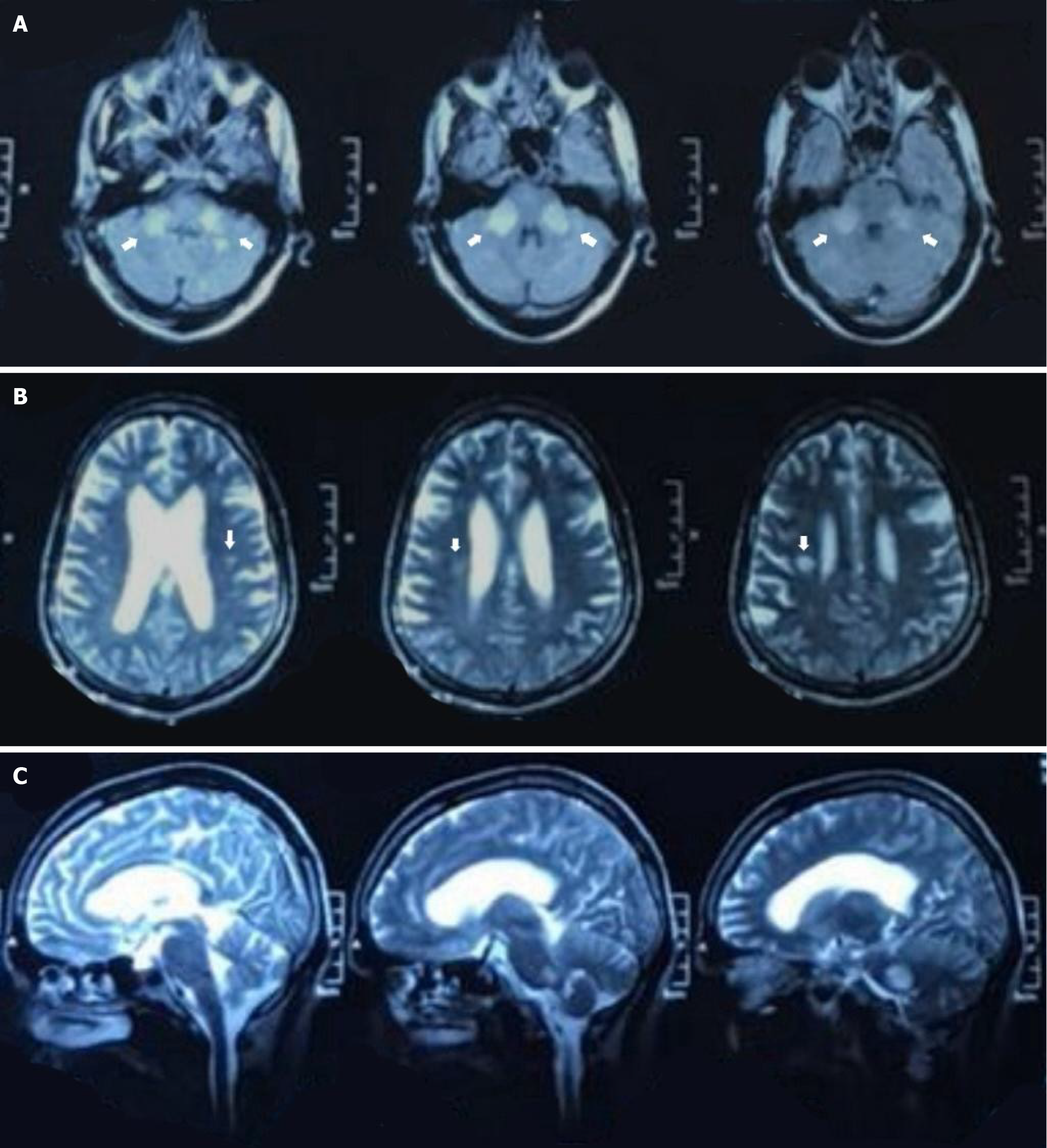
Figure 1 Neuroimaging.
A: Axial T2-weighted magnetic resonance imaging (MRI) demonstrating bilateral, symmetrical hyperintensities in the middle cerebellar peduncles; B: Axial fluid-attenuated inversion recovery MRI image showing subtle periventricular white matter hyperintensities without mass effect or enhancement; C: Sagittal T2-weighted MRI view highlighting lesion distribution involving the brainstem and cerebellum.
- Citation: Faisal A, Tariq Z, Latif RU, Amir S, Basit A, Basil AM. Silent triggers and symmetric peduncles - a rare presentation of adult-onset acute disseminated encephalomyelitis: A case report. World J Clin Cases 2025; 13(30): 112419
- URL: https://www.wjgnet.com/2307-8960/full/v13/i30/112419.htm
- DOI: https://dx.doi.org/10.12998/wjcc.v13.i30.112419


