©The Author(s) 2025.
World J Clin Cases. Oct 26, 2025; 13(30): 110475
Published online Oct 26, 2025. doi: 10.12998/wjcc.v13.i30.110475
Published online Oct 26, 2025. doi: 10.12998/wjcc.v13.i30.110475
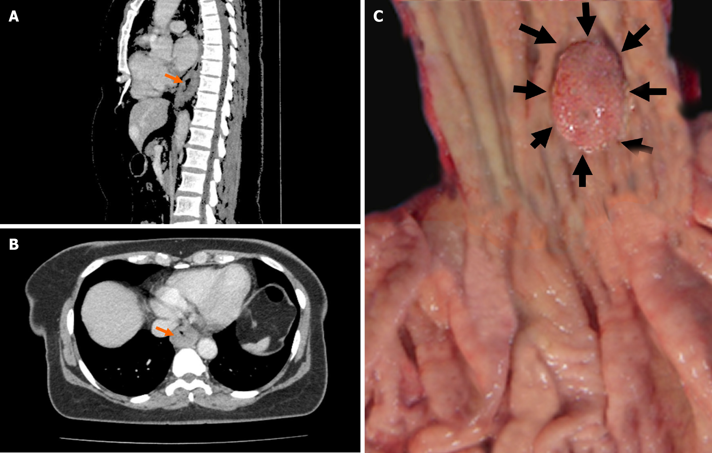
Figure 1 Contrast enhanced computed tomography and macroscopic findings.
A: In the contrast enhanced computed tomography, the sagittal section shows wall thickening in the distal esophagus (orange arrow); B: The axial section reveals wall thickening in the same localization. No lymph node metastasis is observed in either section; C: The macroscopic appearance of the lesion reveals its proximity to the gastroesophageal junction. This location is significant because it can influence both the surgical approach and the overall treatment strategy, given the anatomical and functional importance of the gastroesophageal junction.
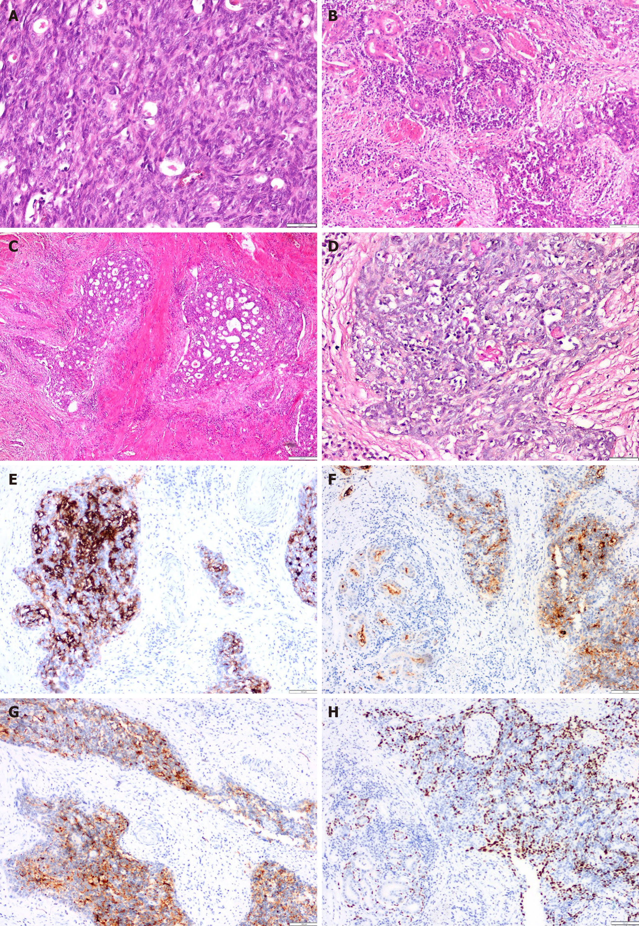
Figure 2 Microscopic appearance of the tumor.
A: The tumor was primarily composed of solid tumor islands. These areas comprised squamous epithelium, intermediate, and adenoid cells with larger cytoplasm and occasional epithelial characteristics; B: Areas displaying gland-like structures (notably in the upper left) were adjacent to solid regions (located in the lower right); C: In another area the glandular structures were interspersed among solid regions, suggesting a mucoepidermoid morphology; D: The mucicarmine staining technique revealed prominent pink staining of mucin (MUC) within the luminal spaces of the glandular structures; E: In these areas, the epithelial cells forming gland-like structures demonstrated MUC1 positivity, contrasting with the surrounding squamous epithelial cells that exhibited no staining; F: Carcinoembryonic antigen positivity was noted in the adenoid areas in both the lumen and the cytoplasm; G: Staining with high molecular weight keratin was positive in poorly differentiated squamous epithelial regions surrounding mucus-containing cells; H: Notably, while p40 positivity was present in the squamous epithelial cells, the mucinous tumor cells remained negative.
- Citation: Yolburun SB, Gunizi OC, Elpek GO. Insights from literature of esophageal mucoepidermoid carcinoma in a female: A rare case report. World J Clin Cases 2025; 13(30): 110475
- URL: https://www.wjgnet.com/2307-8960/full/v13/i30/110475.htm
- DOI: https://dx.doi.org/10.12998/wjcc.v13.i30.110475


