©The Author(s) 2025.
World J Clin Cases. Oct 6, 2025; 13(28): 108710
Published online Oct 6, 2025. doi: 10.12998/wjcc.v13.i28.108710
Published online Oct 6, 2025. doi: 10.12998/wjcc.v13.i28.108710
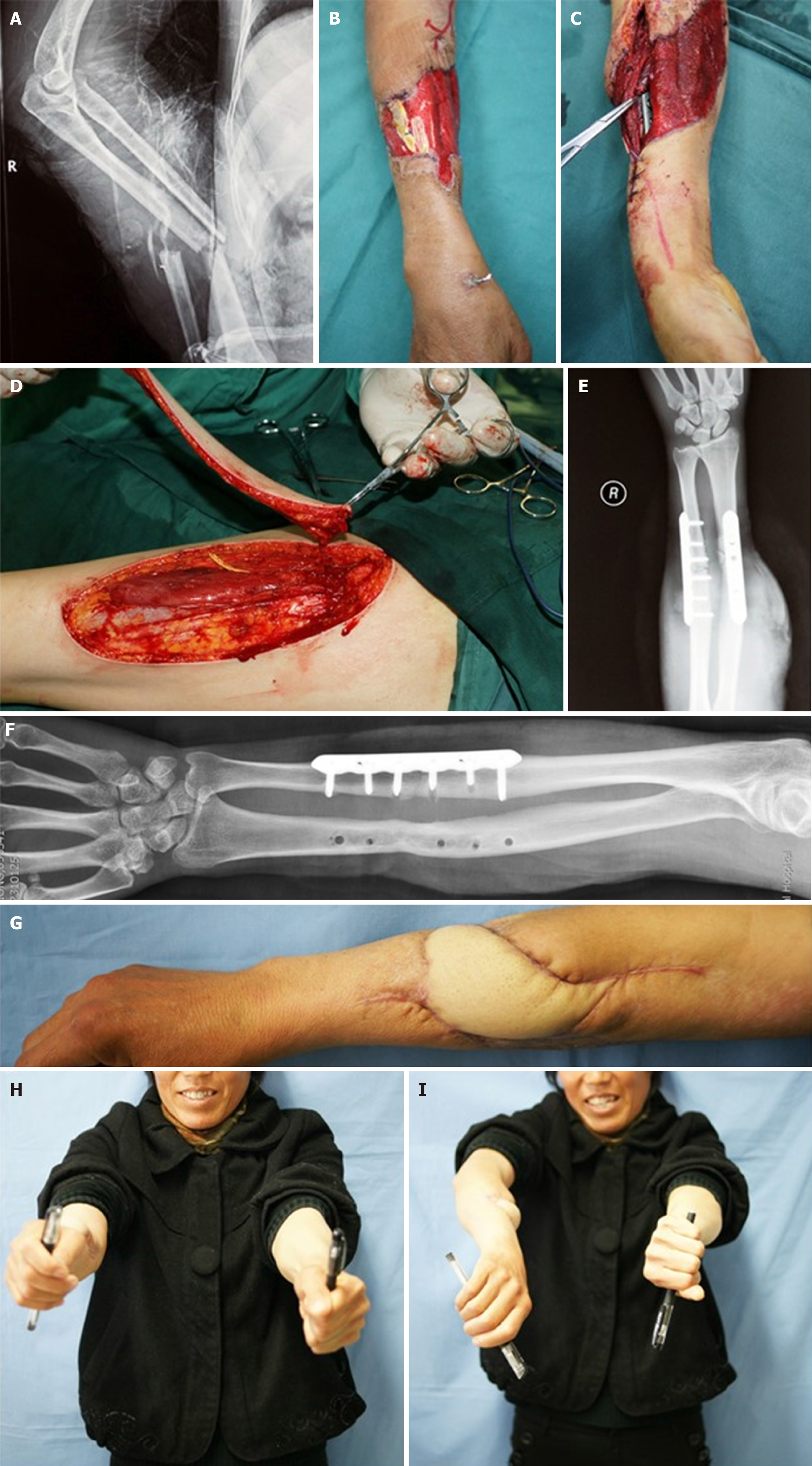
Figure 1 Surgical management of Gustilo IIIB open forearm fracture.
A: Type IIIB open fractures of the forearm; B: Eighteen days after the initial debridement and vacuum-assisted closure; C: Plate fixation; D: Raising of a free lateral circumflex femoral artery perforator flap; E: Anteroposterior (AP) radiograph after surgery; F: Two years post-surgery: AP view; G: Contour of the reconstructed forearm; H and I: Rotation of the forearm compared with the contralateral side.
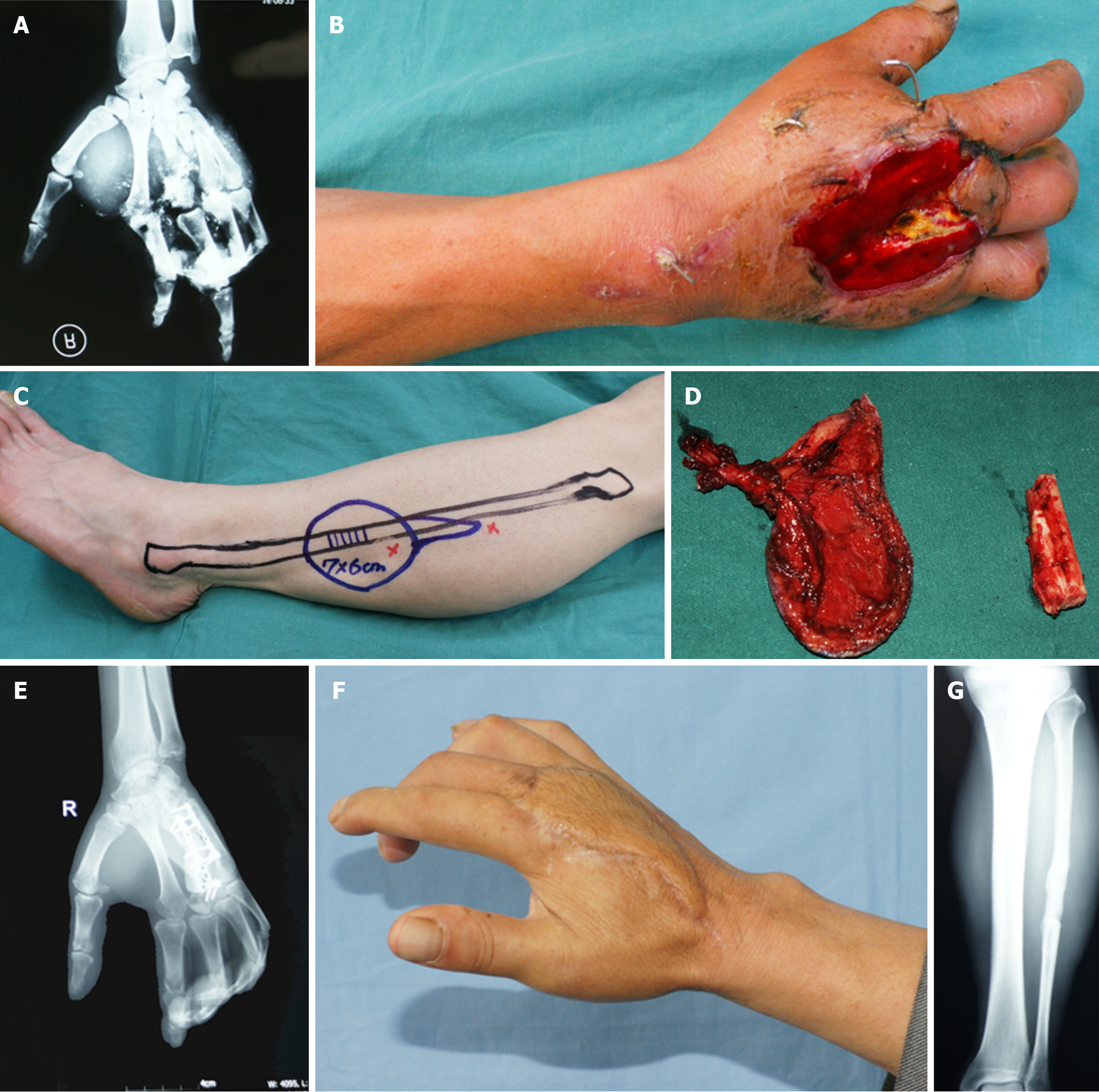
Figure 2 Reconstruction of metacarpal fractures with composite free flap.
A: Open fractures and defects of the 2nd and 3rd metacarpals; B: Wound before definitive surgery; C: Initial design of the composite free flap transfer; D: Free flap and non-vascularized half fibula; E and F: Nine months post-surgery: Bone healing and successful flap integration; G: Re-union of the fibula, minimizing donor site morbidity.
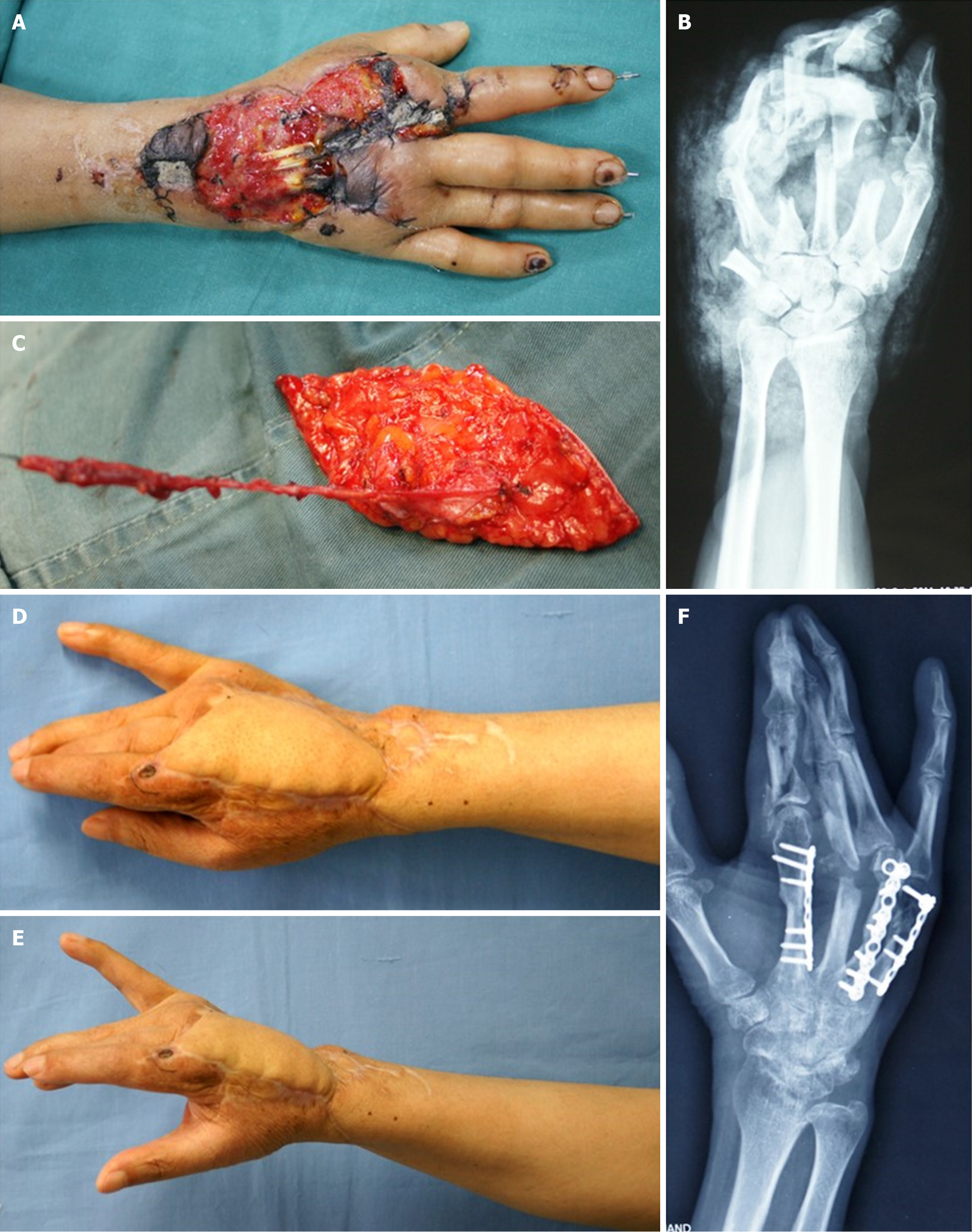
Figure 3 Hand reconstruction with free peroneal artery perforator flap.
A: Open fracture and wound of the hand after repeated debridements; B: Multi-fractured metacarpals; C: Dissected free peroneal artery perforator flap; D and E: Twenty-seven months post-surgery: Flap coverage of the defect and maximal preservation of hand function; F: Bone healing.
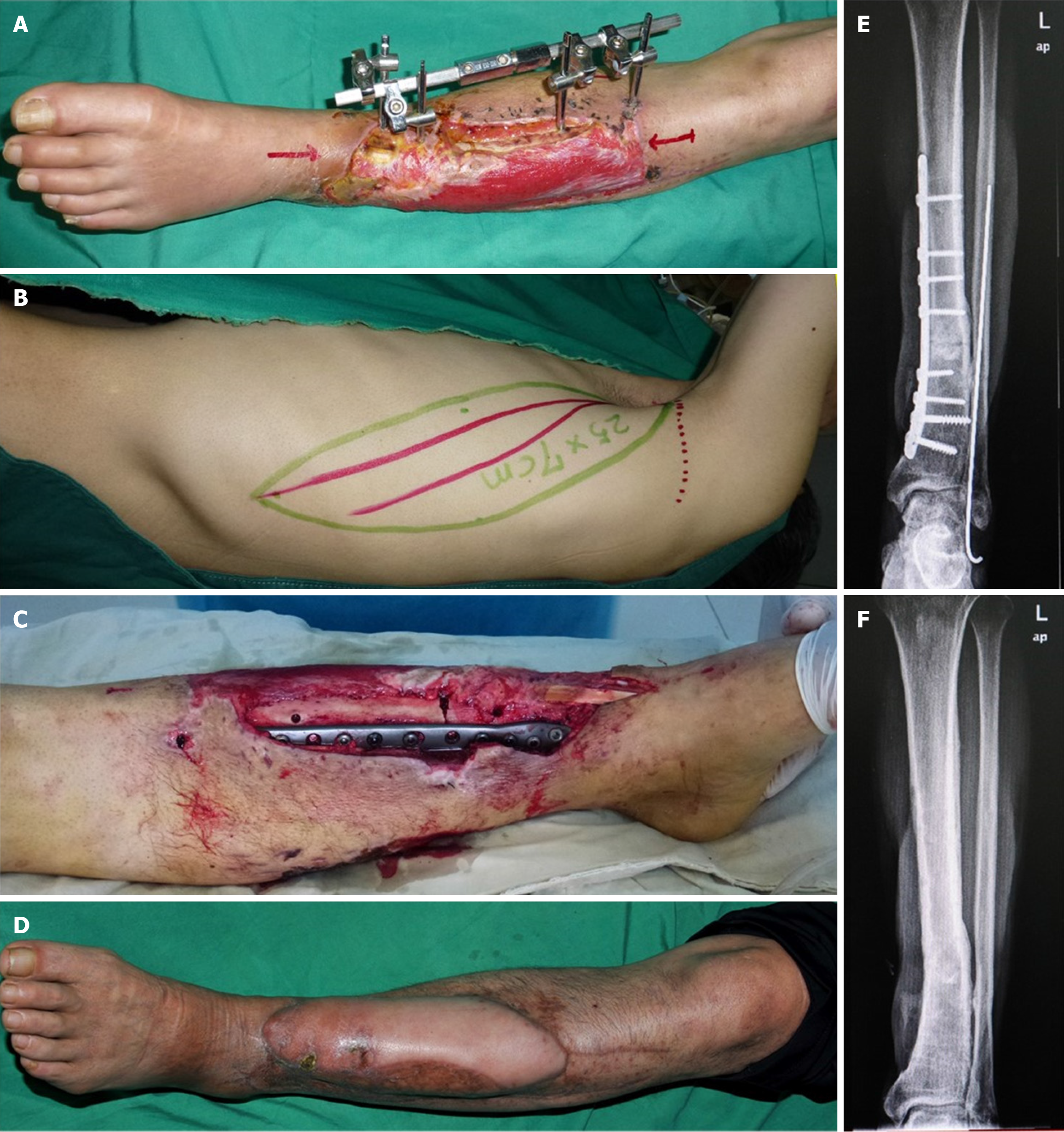
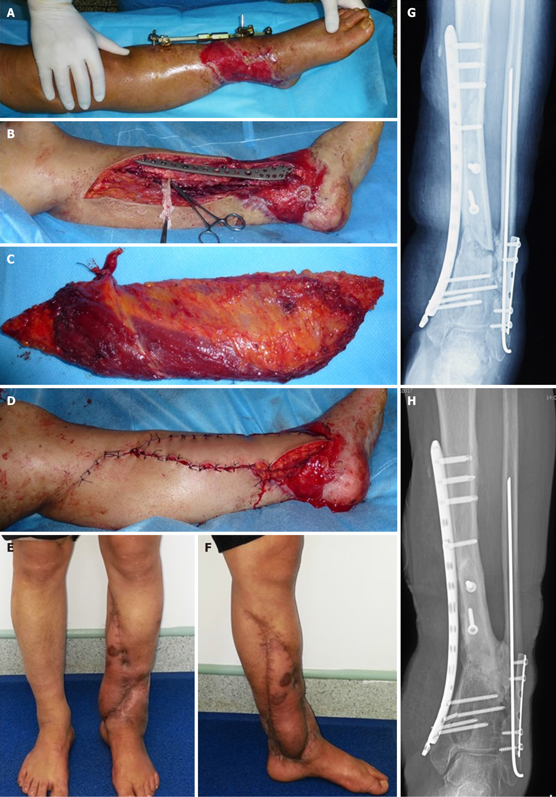
Figure 4 Diabetic lower limb reconstruction with free latissimus dorsi flap.
A: Diabetic leg with open fractures and skin defect; B: Design of the free latissimus dorsi flap; C: Locked compression plate used for bone fixation; D: Appearance of the flap 10 months post-surgery; E: Incomplete bone healing 18 months post-surgery; F: Complete bone healing and removal of internal hardware 30 months post-surgery.
Figure 5 Reconstruction of Gustilo IIIB tibial fracture with free latissimus dorsi flap.
A: Gustilo IIIB fracture of the left leg; B: Removal of external fixation (EF) and fixation with locking compression plates, with exposure of the posterior tibial vessels; C and D: Harvested free latissimus dorsi muscle flap covering both the bone and plate with rich blood circulation; E and F: Thirty months post-surgery: Successful flap integration; G: Bone not united 18 months after surgery; H: Bone healing 29 months post-surgery.
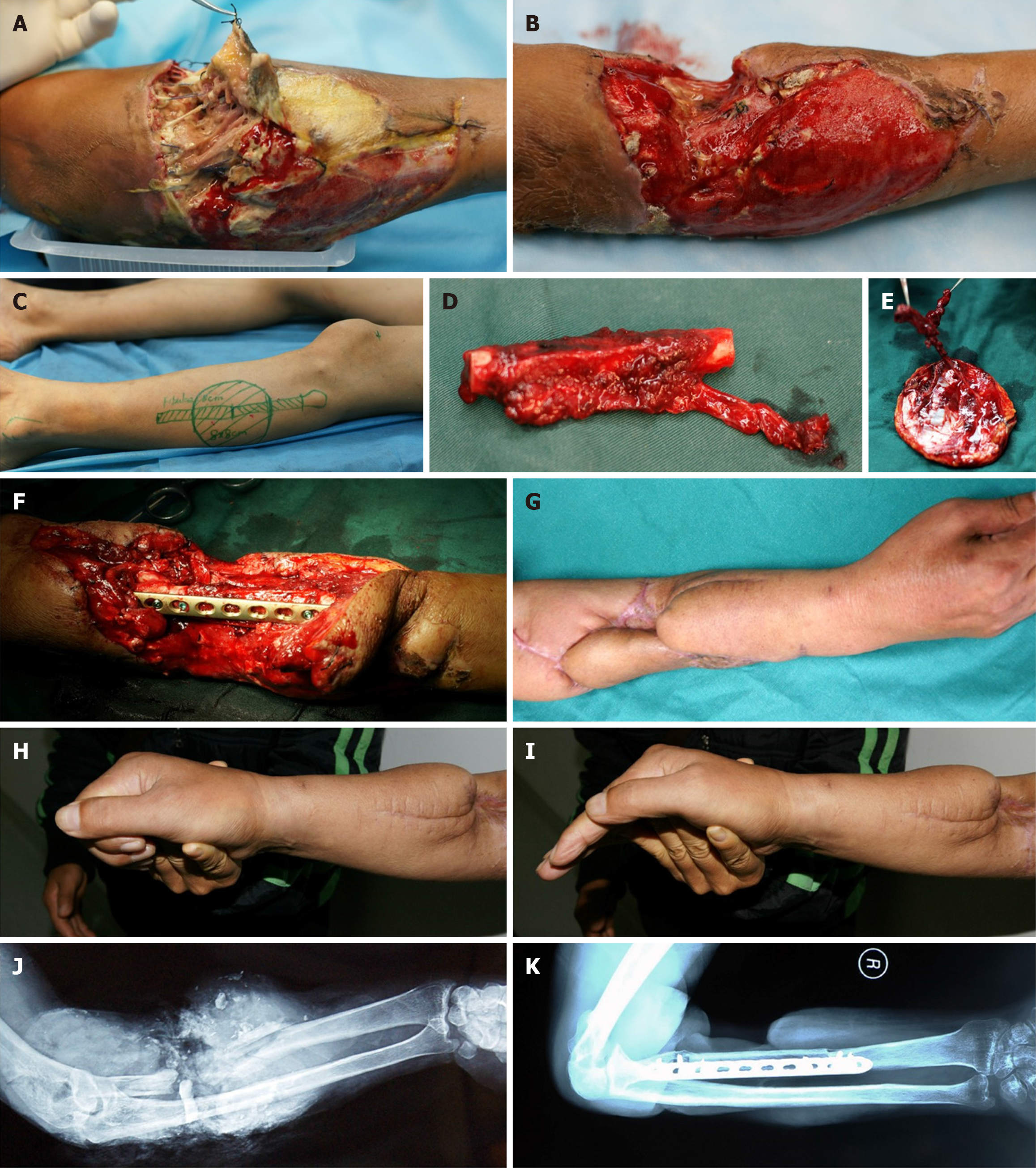
Figure 6 Complex radius reconstruction with composite peroneal flap and free fibula.
A: Gustilo IIIB fracture of the left radius with severe wound infection; B: Dry wound with extensive granulation tissue; C: Design of a composite peroneal perforator flap; D and E: Anastomosis of the free fibula and flap to the recipient site; F: Plate fixation; G-I: Successful flap integration with maximal preservation of wrist function; J and K: Pre- and post-operative radiographs.
- Citation: Bao Q, Pan SW, Gong XK, Wang B, Wei ZN, Xu YQ, Zhu YL, Shi Z. Efficacy of free-flap-transfer and plate-fixation for Gustilo IIIB fractures in type II diabetic patients: A retrospective study. World J Clin Cases 2025; 13(28): 108710
- URL: https://www.wjgnet.com/2307-8960/full/v13/i28/108710.htm
- DOI: https://dx.doi.org/10.12998/wjcc.v13.i28.108710


