©The Author(s) 2025.
World J Clin Cases. Sep 26, 2025; 13(27): 109243
Published online Sep 26, 2025. doi: 10.12998/wjcc.v13.i27.109243
Published online Sep 26, 2025. doi: 10.12998/wjcc.v13.i27.109243
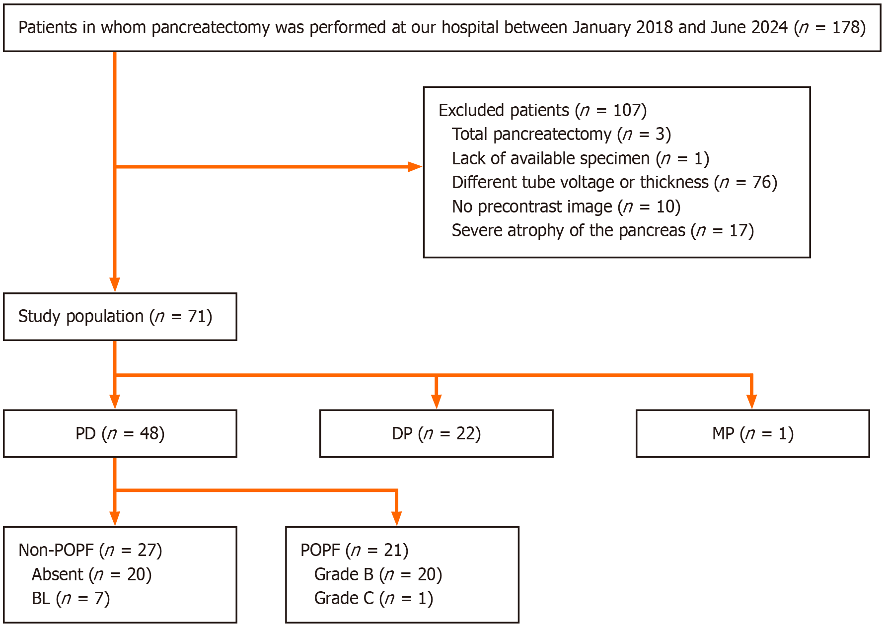
Figure 1 Flow chart of study population.
PD: Pancreaticoduodenectomy; DP: Distal pancreatectomy; MP: Middle pancreatectomy; POPF: Postoperative pancreatic fistula; BL: Biochemical leak.
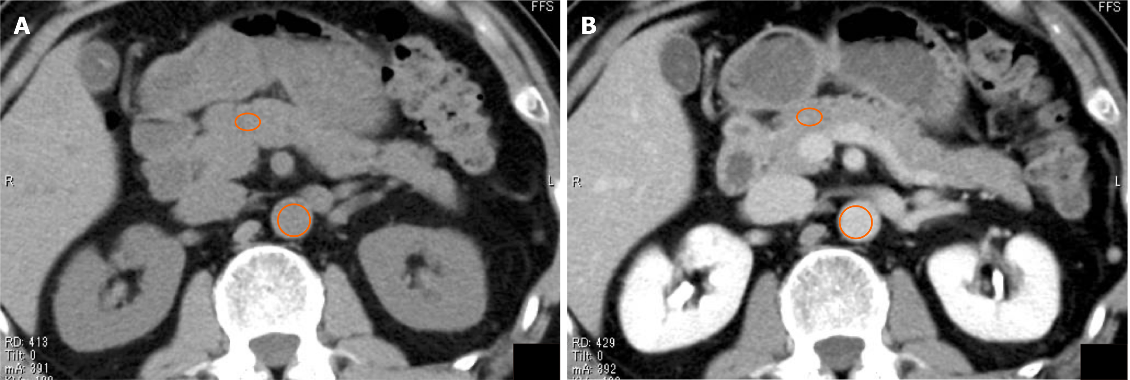
Figure 2 Computed tomography from a 51-year-old male with pancreatic neuroendocrine neoplasm.
A: Represents the precontrast phase; mean computed tomography (CT) value of the pancreas (ellipse), 45.2 HU and mean CT value of the aorta (circle), 43.6 HU; B: Represents the equilibrium phase, with the mean CT value of the pancreas (ellipse) being 78.3 HU and the mean CT value of the aorta (circle) being 125 HU. Extracellular volume fraction in this case was calculated to be 23.3%.
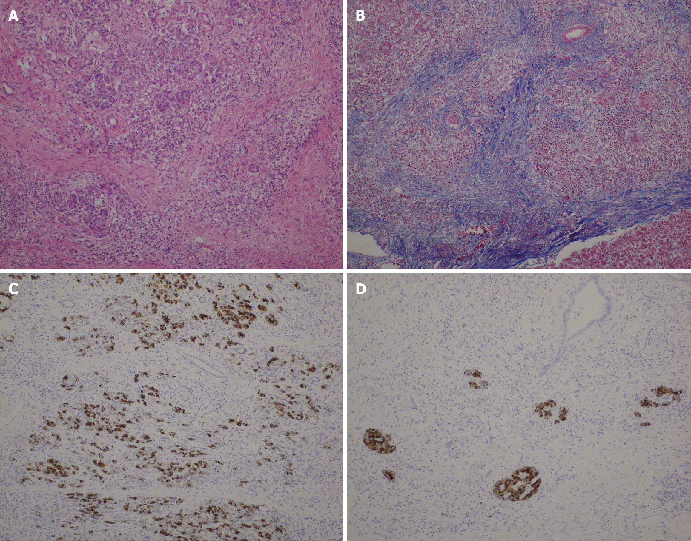
Figure 3 Stained sections from specimens within 1 cm of the pancreatic resection margin.
A: Hematoxylin-eosin staining, 10 × objective lens; B: Masson trichrome staining, 10 × objective lens; C: BCL-10 (333.1) staining, 10 × objective lens; D: Insulin staining, 10 × objective lens.
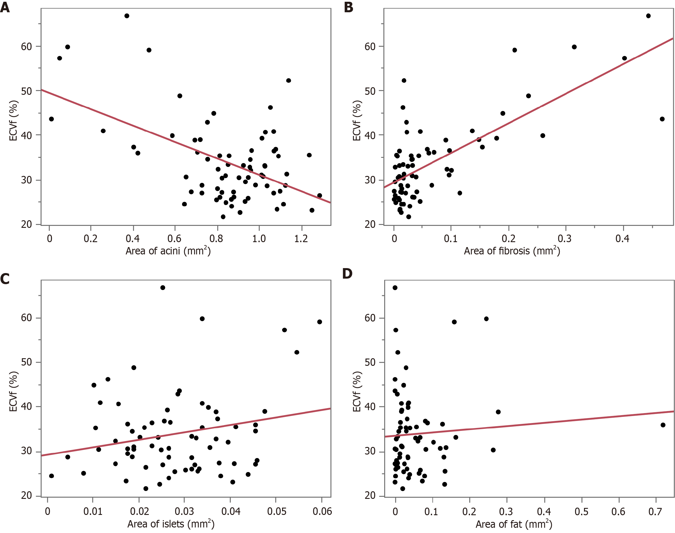
Figure 4 Correlations between extracellular volume fraction and histologic findings.
A: Acini showed a moderate negative correlation with extracellular volume fraction (ECVf) (r = -0.510; P < 0.001); B: Fibrosis showed a strong positive correlation with ECVf (r = 0.724; P < 0.001); C: Islets showed no significant correlation with ECVf (r = 0.228; P = 0.056); D: Fat showed no significant correlation with ECVf (r = 0.075; P = 0.534).
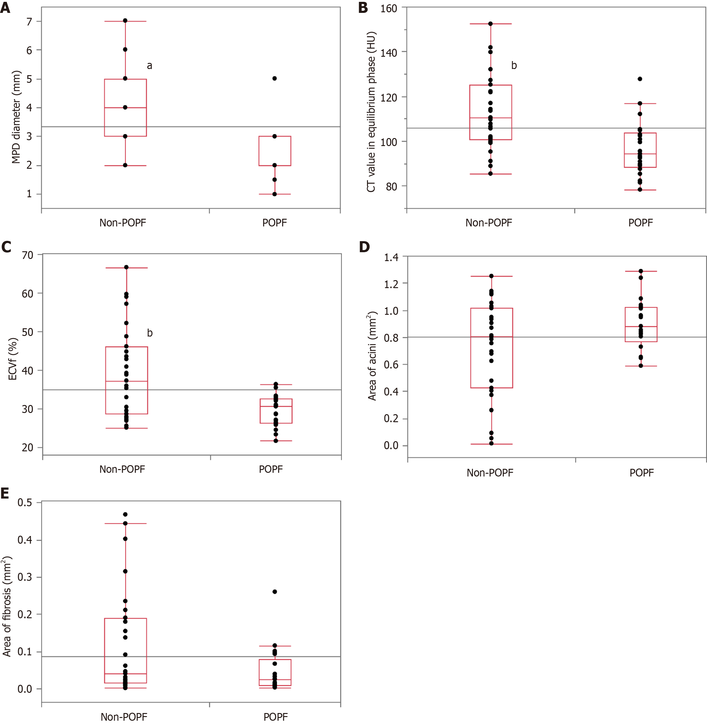
Figure 5 Comparison between non-postoperative pancreatic fistula group and postoperative pancreatic fistula group in the imaging and histopathological findings.
A: Main pancreatic duct diameter (4 mm vs 2 mm; P < 0.001); B: Computed tomography value in equilibrium phase (110.5 HU vs 94.3 HU; P = 0.001); C: Extracellular volume fraction (37.2% vs 30.5%; P = 0.003); D: Area of acini (0.802 mm2vs 0.880 mm2; P = 0.127); E: Area of fibrosis (0.041 mm² vs 0.026 mm²; P = 0.110). aP < 0.001; bP < 0.01. POPF: Postoperative pancreatic fistula; MPD: Main pancreatic duct; CT: Computed tomography; ECVf: Extracellular volume fraction.
- Citation: Nakamura A, Ogawa T, Tanaka K, Takahashi Y, Murai S, Tashiro Y, Wada A, Ueda Y, Sasaki Y, Minegishi Y, Matsuo K, Yamochi T. Estimation of pancreatic histology and likelihood of postoperative pancreatic fistula using extracellular volume fraction from contrast-enhanced computed tomography. World J Clin Cases 2025; 13(27): 109243
- URL: https://www.wjgnet.com/2307-8960/full/v13/i27/109243.htm
- DOI: https://dx.doi.org/10.12998/wjcc.v13.i27.109243


