©The Author(s) 2025.
World J Clin Cases. Sep 26, 2025; 13(27): 108998
Published online Sep 26, 2025. doi: 10.12998/wjcc.v13.i27.108998
Published online Sep 26, 2025. doi: 10.12998/wjcc.v13.i27.108998
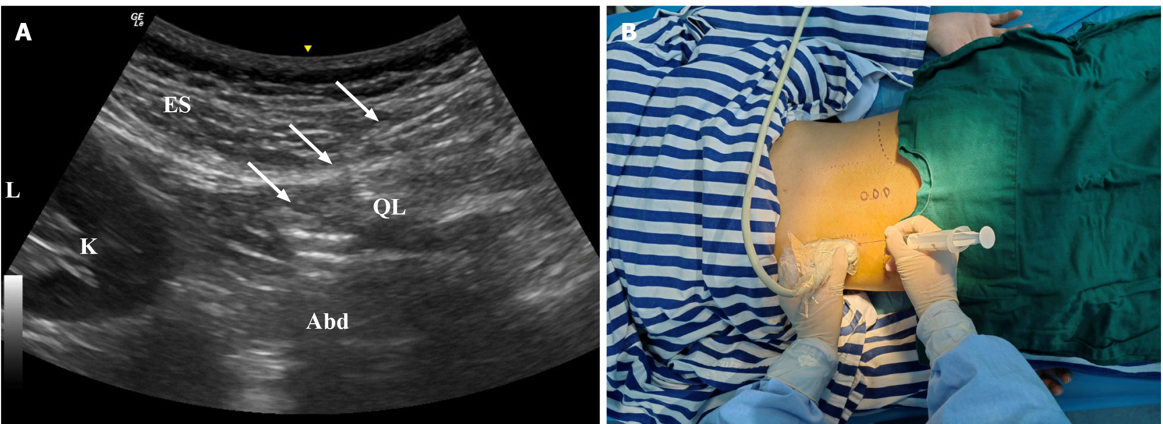
Figure 1 Under the guidance of ultrasound, the doctor precisely injected platelet-rich plasma into the quadratus lumborum of the individual.
A: Ultrasound; B: Injected platelet-rich plasma. White arrows indicate the needle trajectory. L: Left; ES: Erector spinae; QL: Quadratus lumborum; K: Kidney; Abd: Abdomen.
Figure 2 Longitudinal assessment of lumbar erector spinae muscle symmetry following ultrasound-guided platelet-rich plasma therapy.
A: Pretreatment measurement demonstrating significant asymmetry (left: 2.3 cm; right: 3.6 cm; difference: 1.3 cm); B: 3-month post-treatment reduction in asymmetry (left: 3.5 cm; right: 3.8 cm; difference: 0.3 cm); C: 6-month follow-up showing near-symmetrical restoration (left: 4.4 cm; right: 4.6 cm; difference: 0.2 cm). White arrow: Affected side (left erector spinae). Black arrow: Unaffected side (right erector spinae). Dashed line: Muscle width measurement boundaries. Circle: Palpated spinous process landmark. L: Left side.
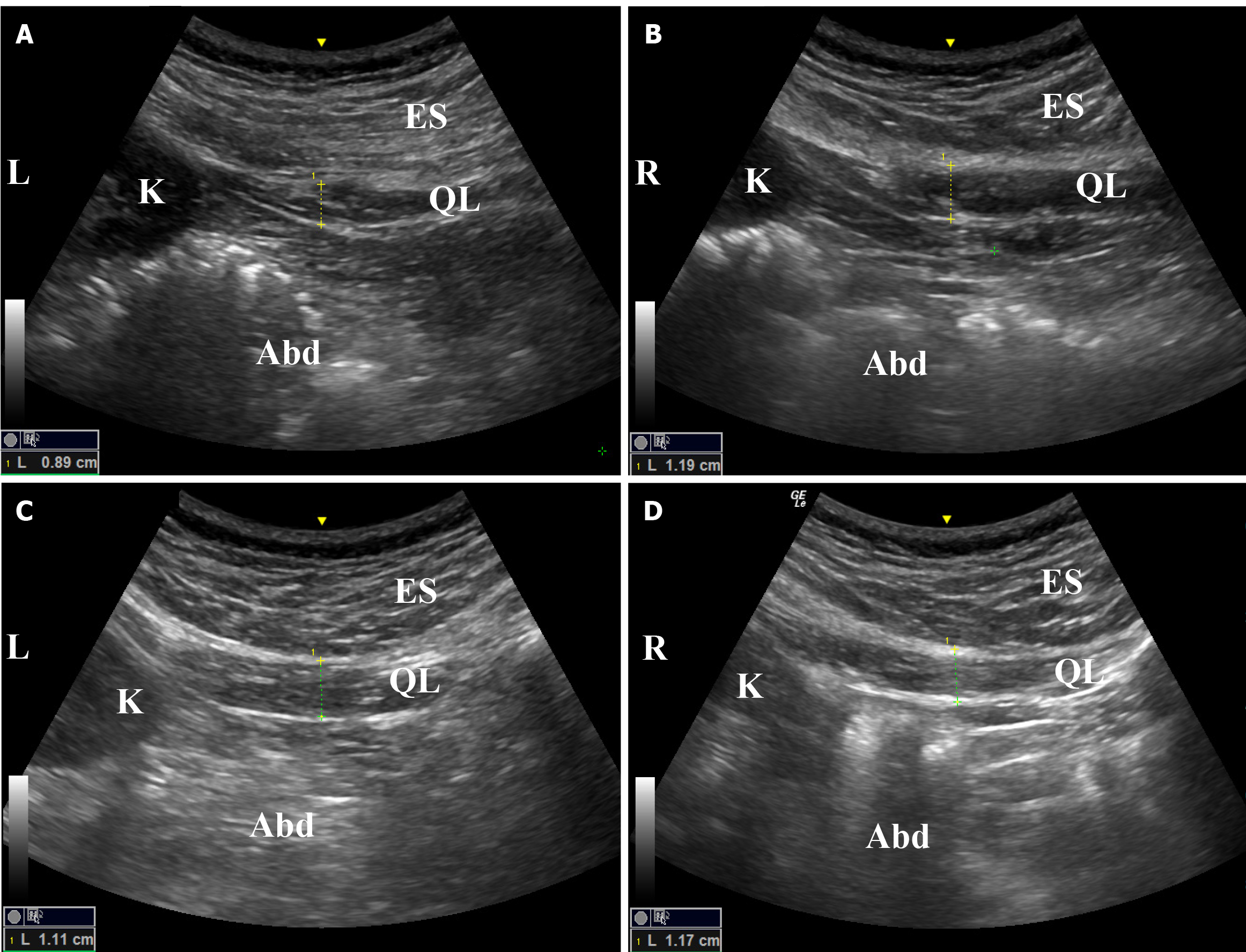
Figure 3 After 3-month of treatment, the difference in the muscle thickness measurements of bilateral quadratus lumborum was reduced from 0.
3 to 0.06 cm in ultrasound examination. A: Before treatment left side; B: Before treatment right side; C: 3 months after treatment left side; D: 3 months after treatment right side. L: Left; R: Right; ES: Erector spinae; QL: Quadratus lumborum; K: Kidney; Abd: Abdomen.
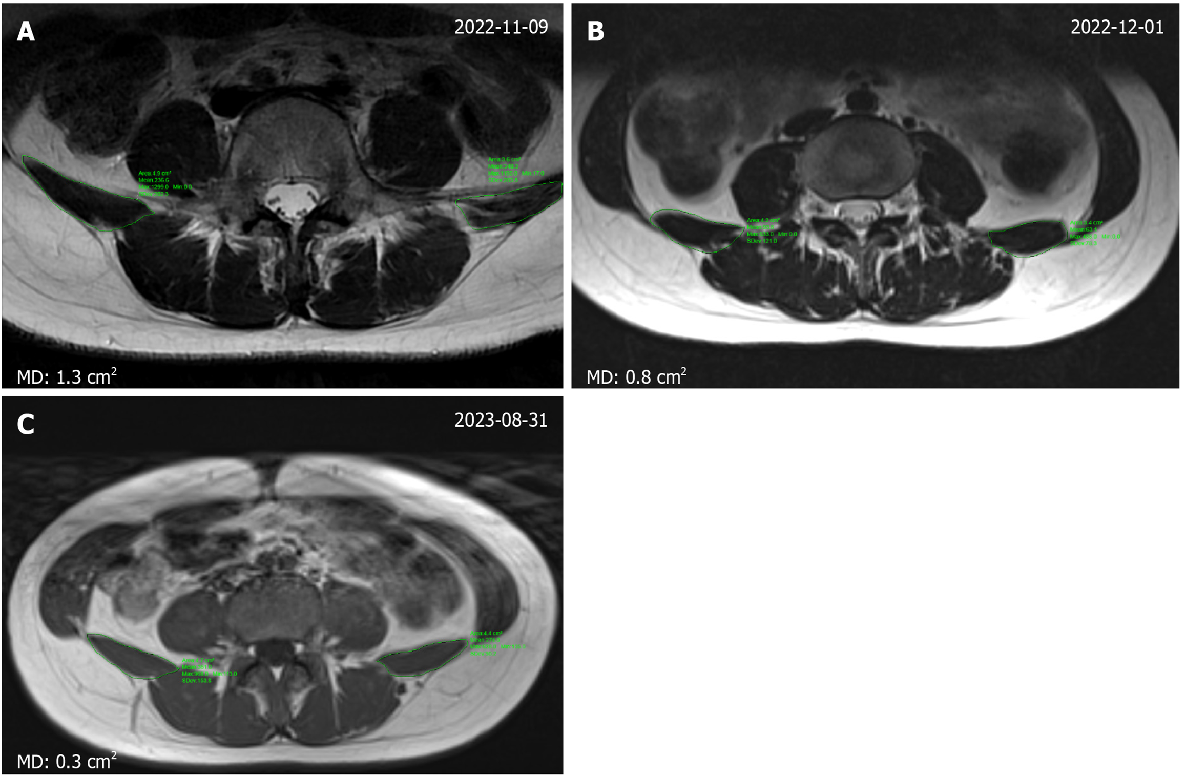
Figure 4 Magnetic resonance imaging.
A: Mean difference of the cross-sectional area of the right and left quadratus lumborum measured by magnetic resonance imaging before treatment was 1.3 cm2; B: At 1-month follow-up, the mean difference of the right and left side (MD0 between the left and right was reduced to 0.8 cm2; C: In the last follow-up after 10 month of treatment, the MD between the left and right sides was further reduced to 0.3 cm2; The symmetry of both sides of muscle dimension gradually restored. MD: Mean difference of the right and left side.
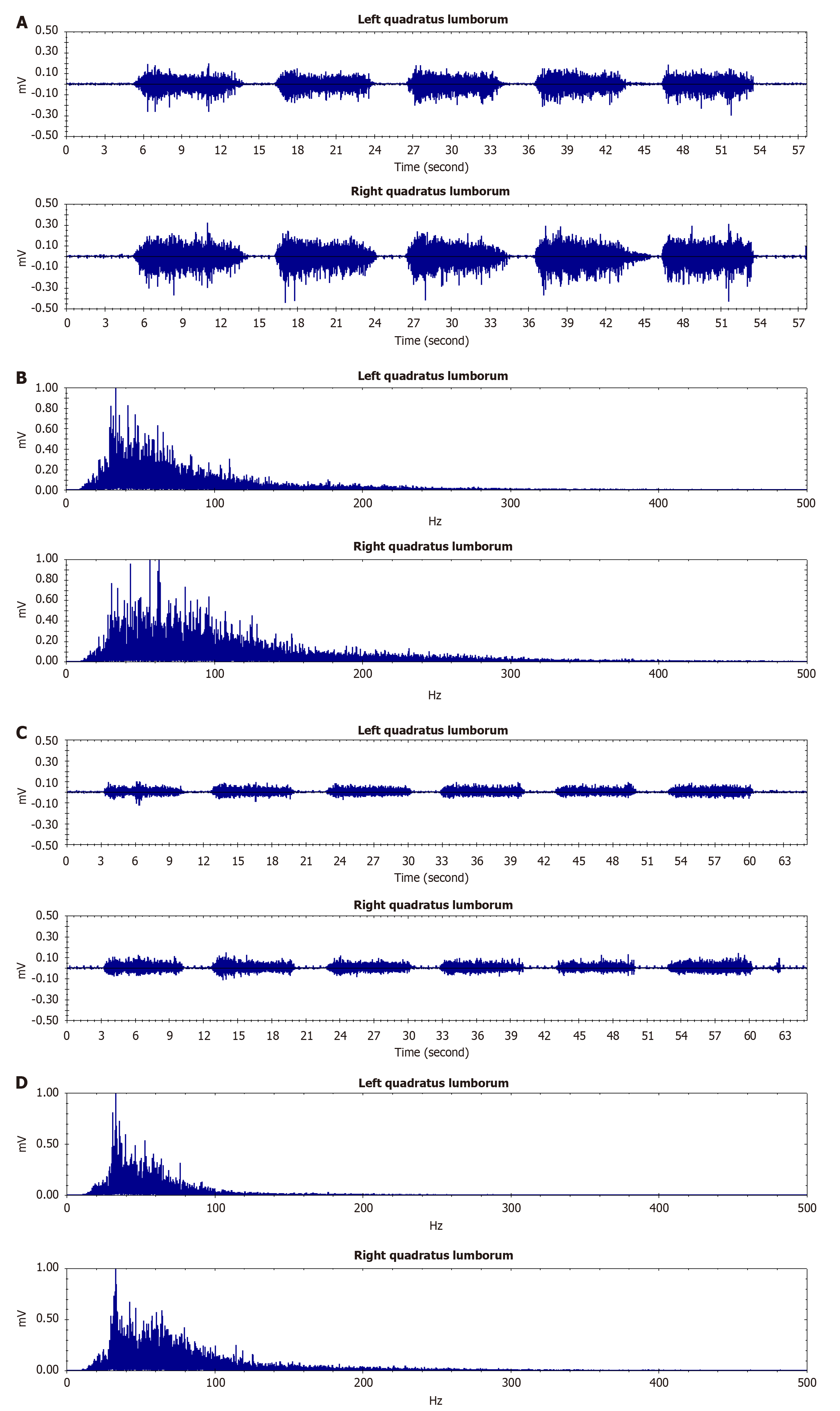
Figure 5 The surface electromyography of the bilateral quadratus lumborum revealed improved symmetry in both time-domain and frequency-domain analyses following platelet-rich plasma treatment compared to pretreatment measurements.
The frequency-domain analysis demonstrated a significant reduction in muscle fatigue at the 6-month follow-up assessment. A: Surface electromyography (sEMG) time domain analysis of quadratus lumborum (QL) before treatment; B: sEMG frequency domain analysis of QL before treatment; C: sEMG time domain analysis of QL at 6-month follow-up after treatment; D: sEMG frequency domain analysis of QL at 6-month follow-up after treatment.
- Citation: Ai SL, Xiang XN, Yu X, Li N, Zhang XY, Zhang KB, Jiang HY, Wang Q, He HC. Ultrasound-guided platelet-rich plasma injection improves pain, function and symmetry in lumbar myofascial pain syndrome: A case report. World J Clin Cases 2025; 13(27): 108998
- URL: https://www.wjgnet.com/2307-8960/full/v13/i27/108998.htm
- DOI: https://dx.doi.org/10.12998/wjcc.v13.i27.108998


