©The Author(s) 2025.
World J Clin Cases. Jun 26, 2025; 13(18): 100610
Published online Jun 26, 2025. doi: 10.12998/wjcc.v13.i18.100610
Published online Jun 26, 2025. doi: 10.12998/wjcc.v13.i18.100610
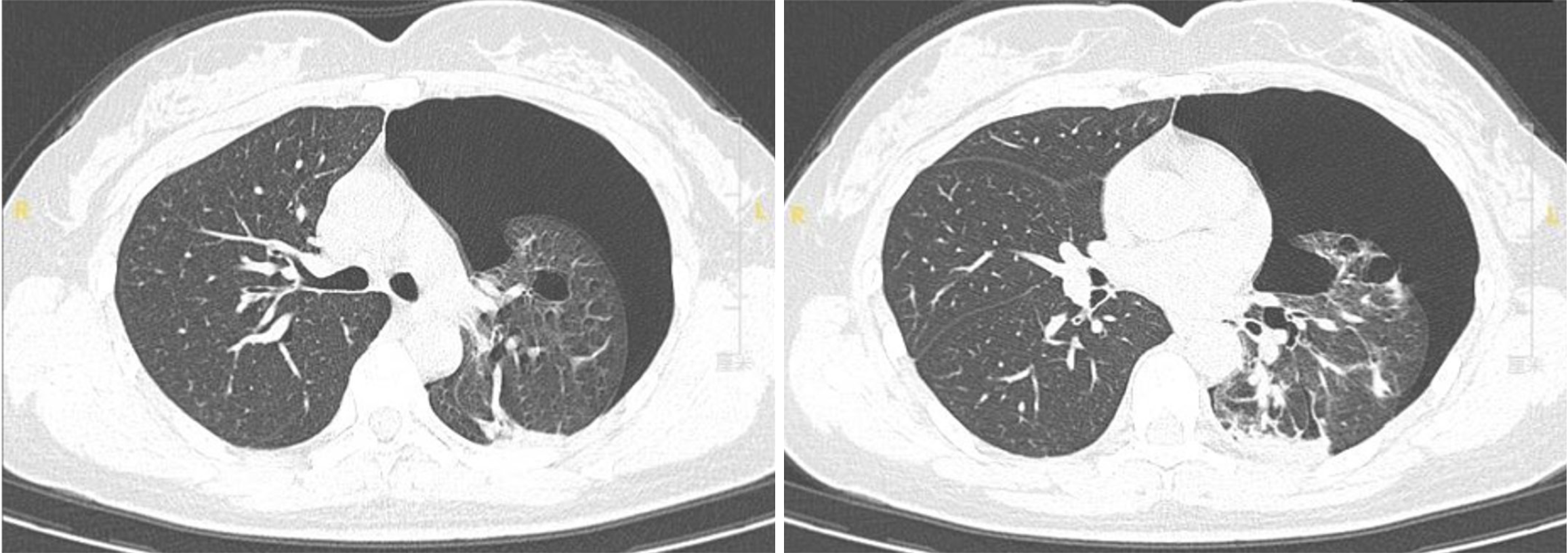
Figure 1 A chest computed tomography scan revealing left-sided pneumothorax and multiple thin-walled cysts.
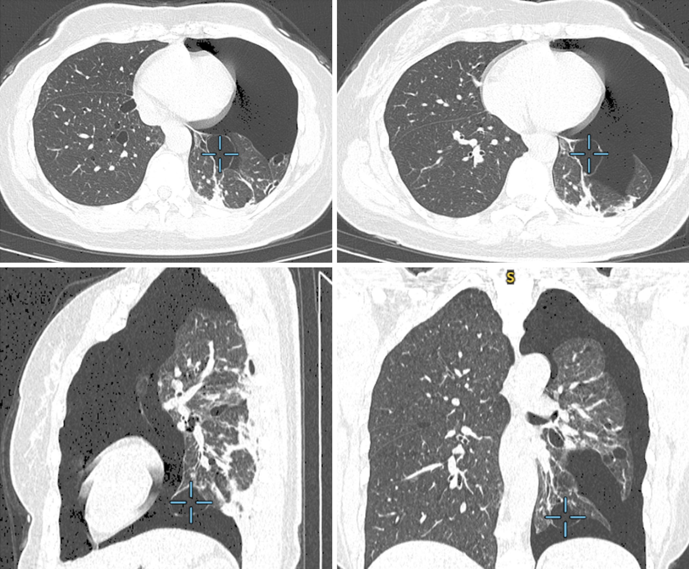
Figure 2 Chest computed tomography volume reconstruction showing pneumothorax and multiple pulmonary cysts.
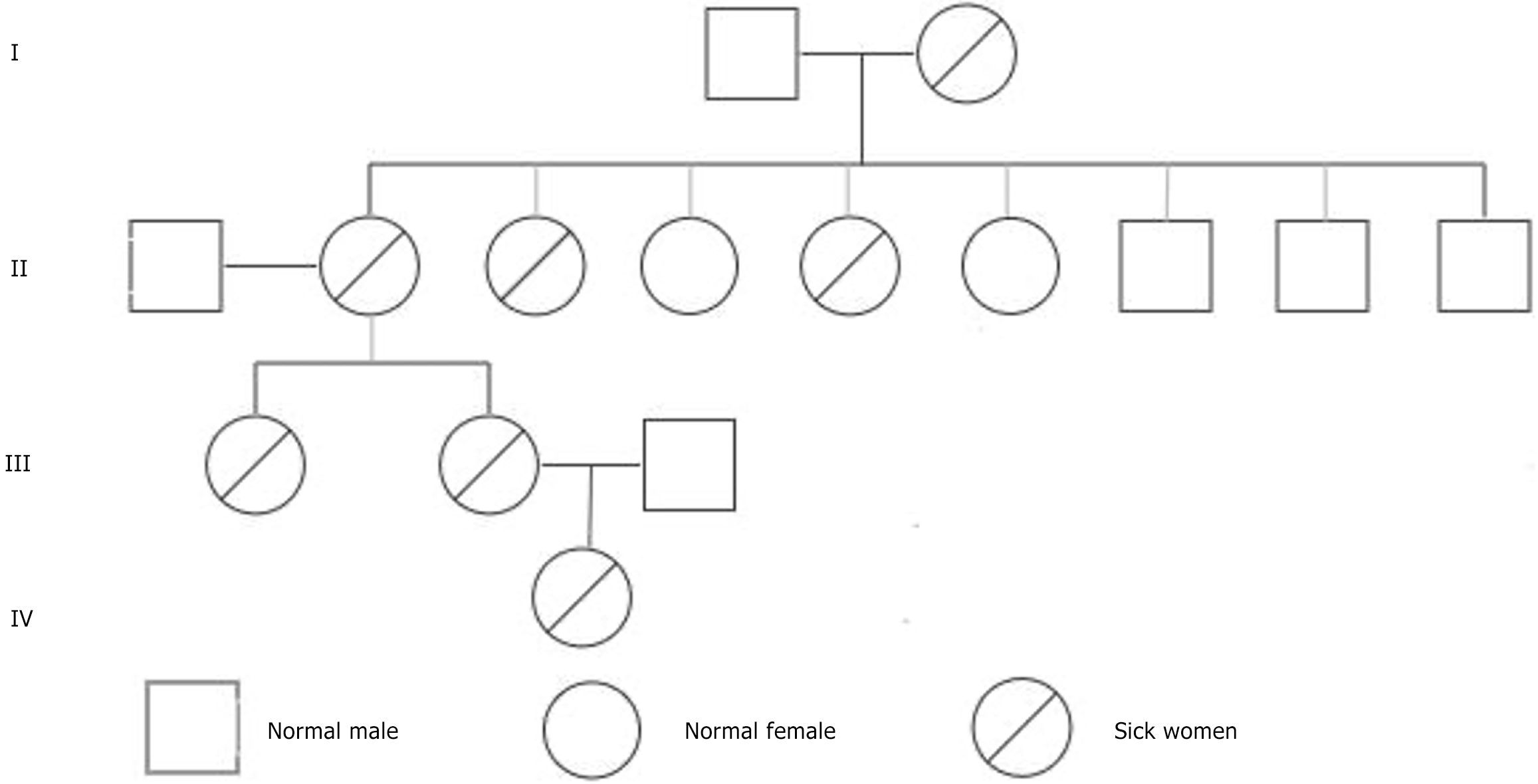
Figure 3 Medical conditions of the patient's family members.
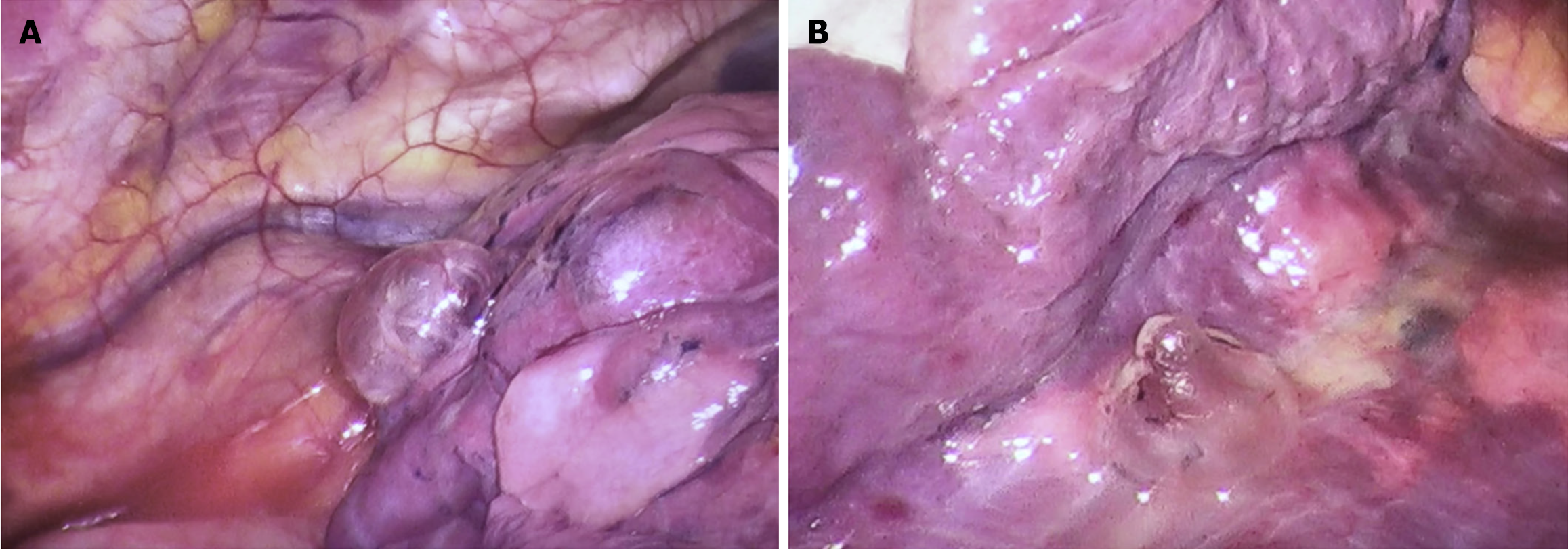
Figure 4 Multiple bullae in the thoracic cavity were observed under intraoperative thoracoscopy.
A: Multiple bullae in the apical segment of the left upper lobe, presenting as fish bubble-like structures; B: Bullae near the diaphragmatic surface of the left lower lobe, exhibiting a fish hook-shaped appearance.
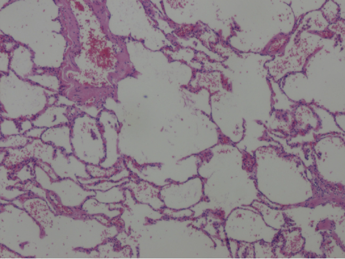
Figure 5 Postoperative pathological findings.
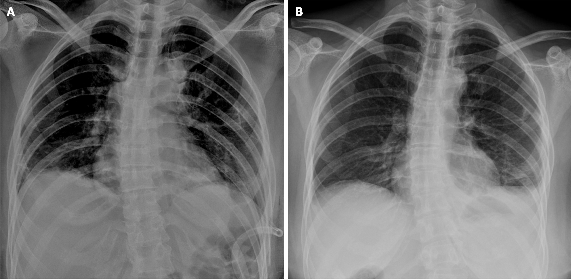
Figure 6 Postoperative review results of the patient.
A: The first day after the surgery; B: One month after the surgery.
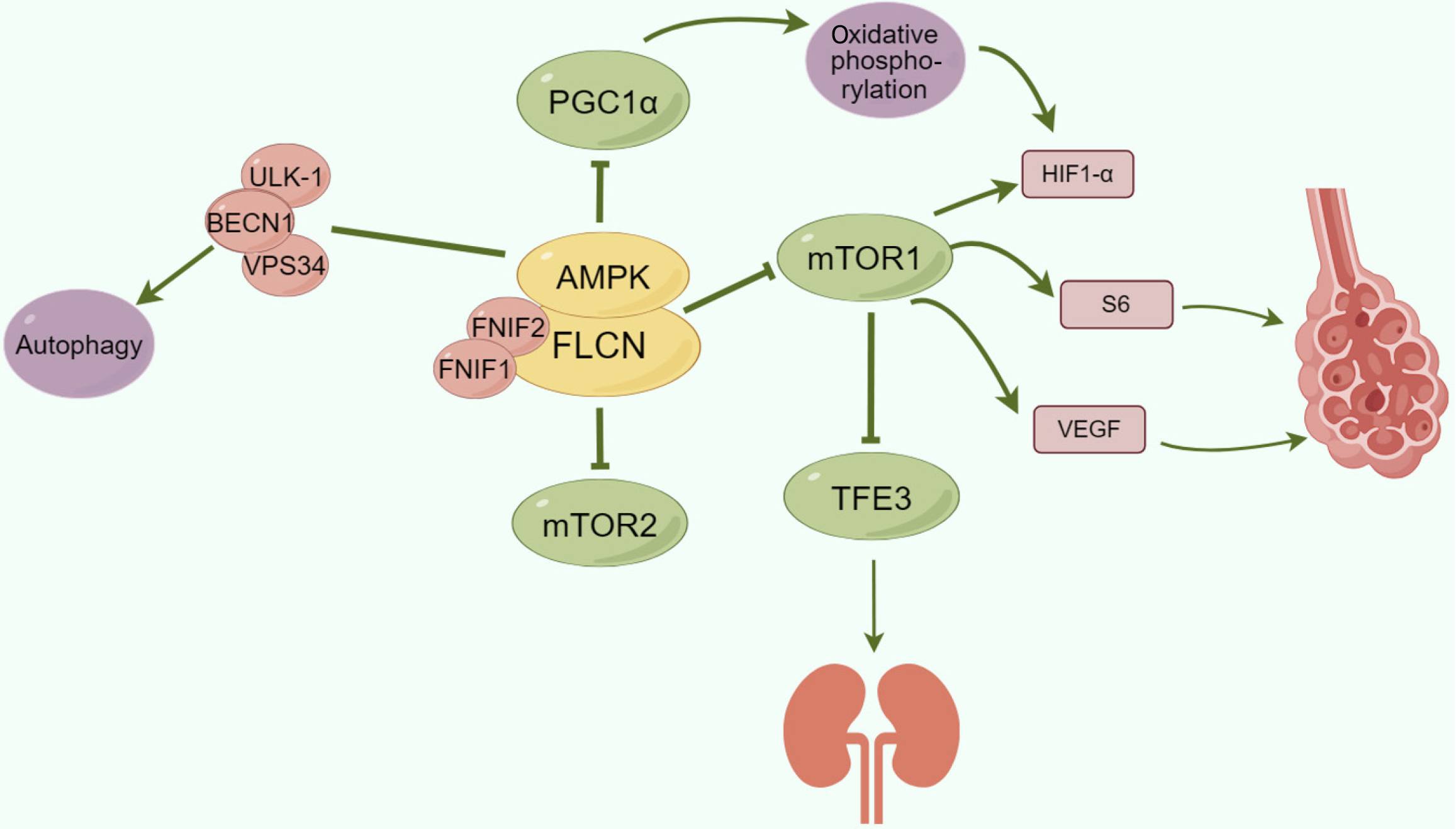
Figure 7 FLCN-associated pathways.
- Citation: Li MZ, Deng J. Birt-Hogg-Dubé syndrome - a rare genetic disorder complicated by pneumothorax: A case report. World J Clin Cases 2025; 13(18): 100610
- URL: https://www.wjgnet.com/2307-8960/full/v13/i18/100610.htm
- DOI: https://dx.doi.org/10.12998/wjcc.v13.i18.100610


