©The Author(s) 2024.
World J Clin Cases. Feb 26, 2024; 12(6): 1076-1083
Published online Feb 26, 2024. doi: 10.12998/wjcc.v12.i6.1076
Published online Feb 26, 2024. doi: 10.12998/wjcc.v12.i6.1076
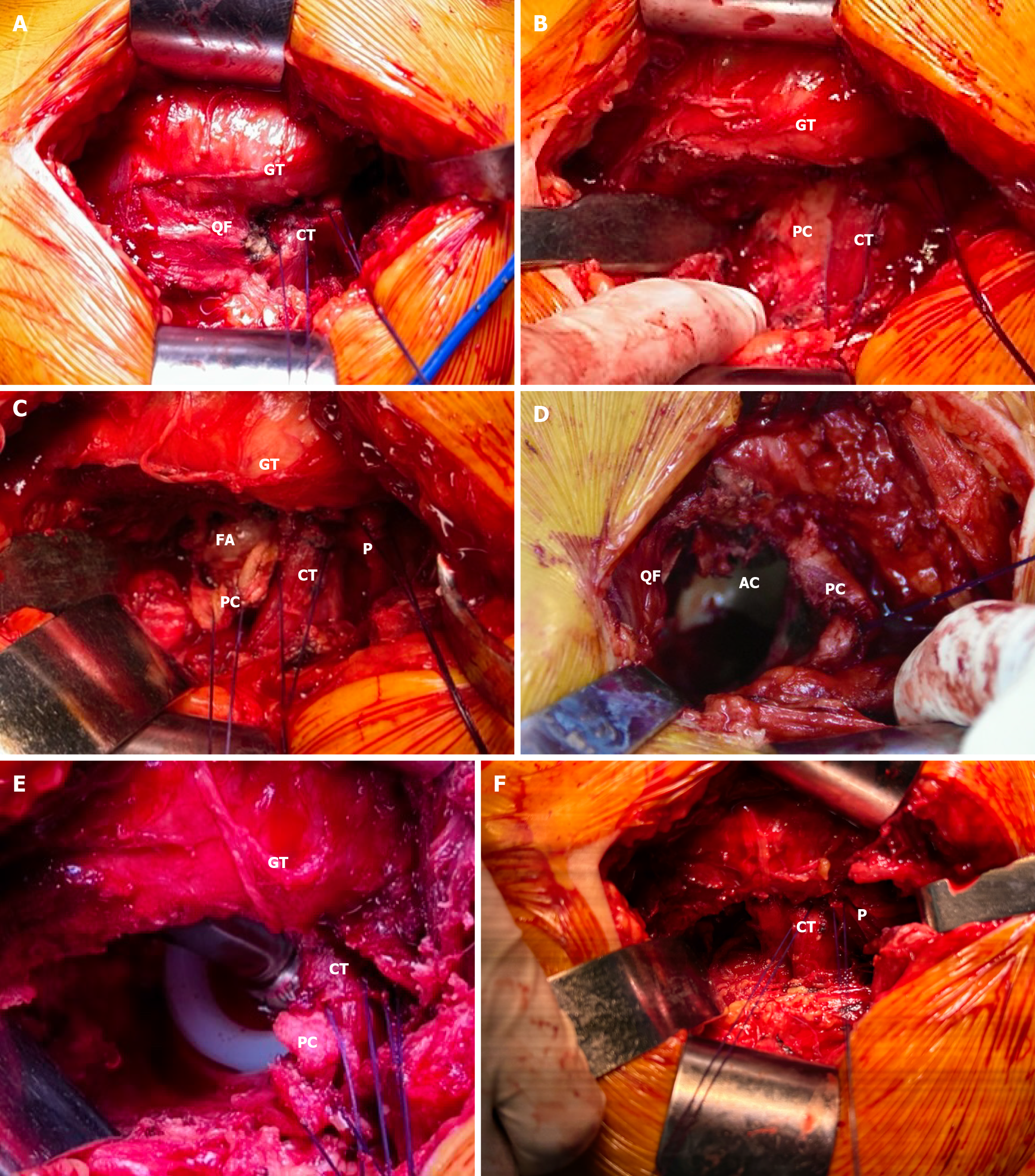
Figure 1 Shows the exposure of left hip joint, protection of the external external rotator muscles and piriformis tendon during bipolar hip arthroplasty using conjoined tendon preserving posterior lateral approach.
A: After self-retaining retractors were placed under the deep fascia layer of the gluteus maximus, the piriformis and conjoined tendon were marked by two No. 1 Ethicon sutures. Expose the greater trochanter and quadratus femoris muscle; B: The partial quadratus femoris muscle was incised and retracted by a Hohmann retractor. The posterior capsule was exposed; C: The bottom half of the posterior capsule was incised and inverted by two sutures. Expose the femoral head D: After removing the femoral head, the acetabulum was exposed. A Hohmann retractor was placed behind the posterior-inferior wall to better observe the acetabulum; E: The prosthesis was implanted, and the marking sutures remained in place; F: The posterior capsule was reattached at the trochanteric insertion site. The conjoined tendon and piriformis marked by sutures remained intact. GT: Greater trochanter; CT: Conjoined tendon; PC: Posterior capsule; FA: Femoral head; QF: Quadratus femoris muscle; AC: Acetabulum; P: Piriformis.
- Citation: Yan TX, Dong SJ, Ning B, Zhao YC. Bipolar hip arthroplasty using conjoined tendon preserving posterior lateral approach in treatment of displaced femoral neck fractures. World J Clin Cases 2024; 12(6): 1076-1083
- URL: https://www.wjgnet.com/2307-8960/full/v12/i6/1076.htm
- DOI: https://dx.doi.org/10.12998/wjcc.v12.i6.1076


