Copyright
©The Author(s) 2024.
World J Clin Cases. Oct 26, 2024; 12(30): 6383-6390
Published online Oct 26, 2024. doi: 10.12998/wjcc.v12.i30.6383
Published online Oct 26, 2024. doi: 10.12998/wjcc.v12.i30.6383
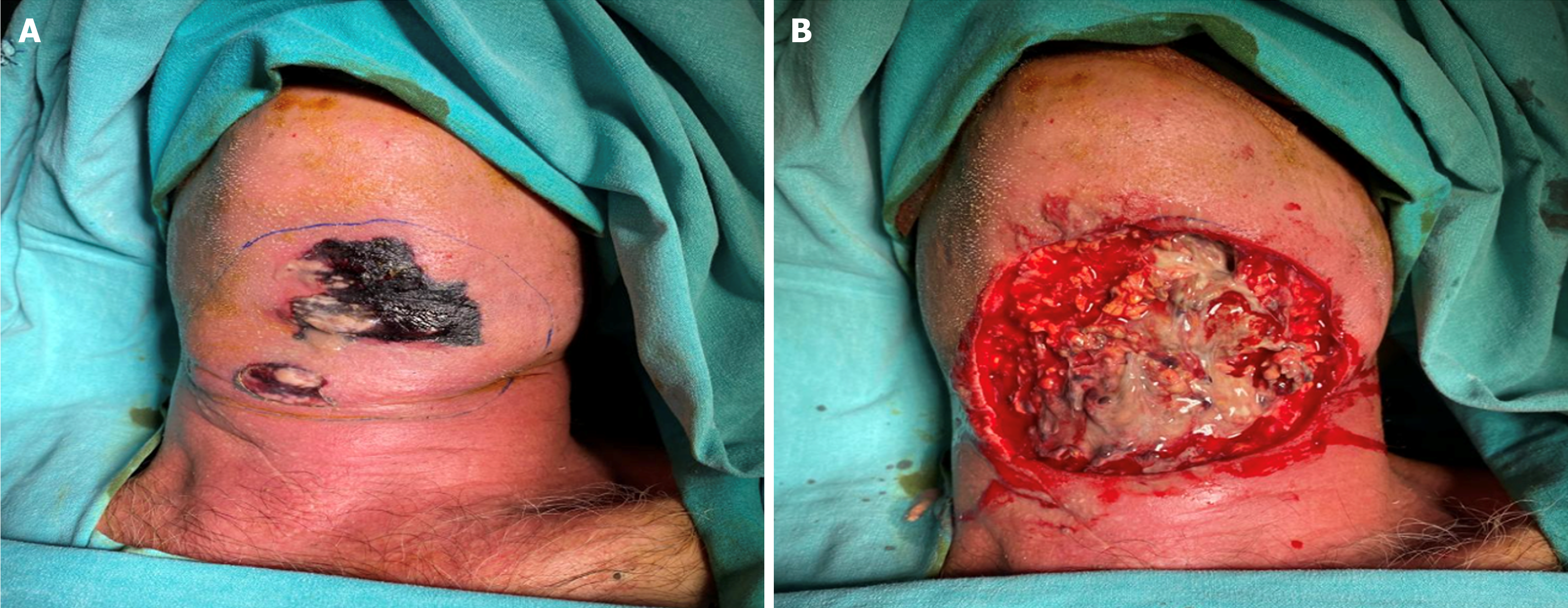
Figure 1 Image of necrotising fasciitis patient.
A: Preoperative image; B: Intraoperative image.
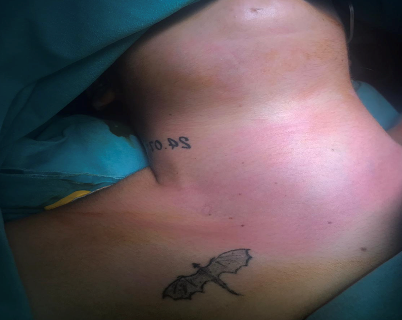
Figure 2
Neck swelling and hyperemia in a deep neck infection patient.
Figure 3 Contrast enhanced neck and thorax computed tomography scans in axial plane.
A: Submandibular and parapharyngeal abscess; B: Cervical necrotising fasciitis; C: Mediastinitis and pleural effusion.
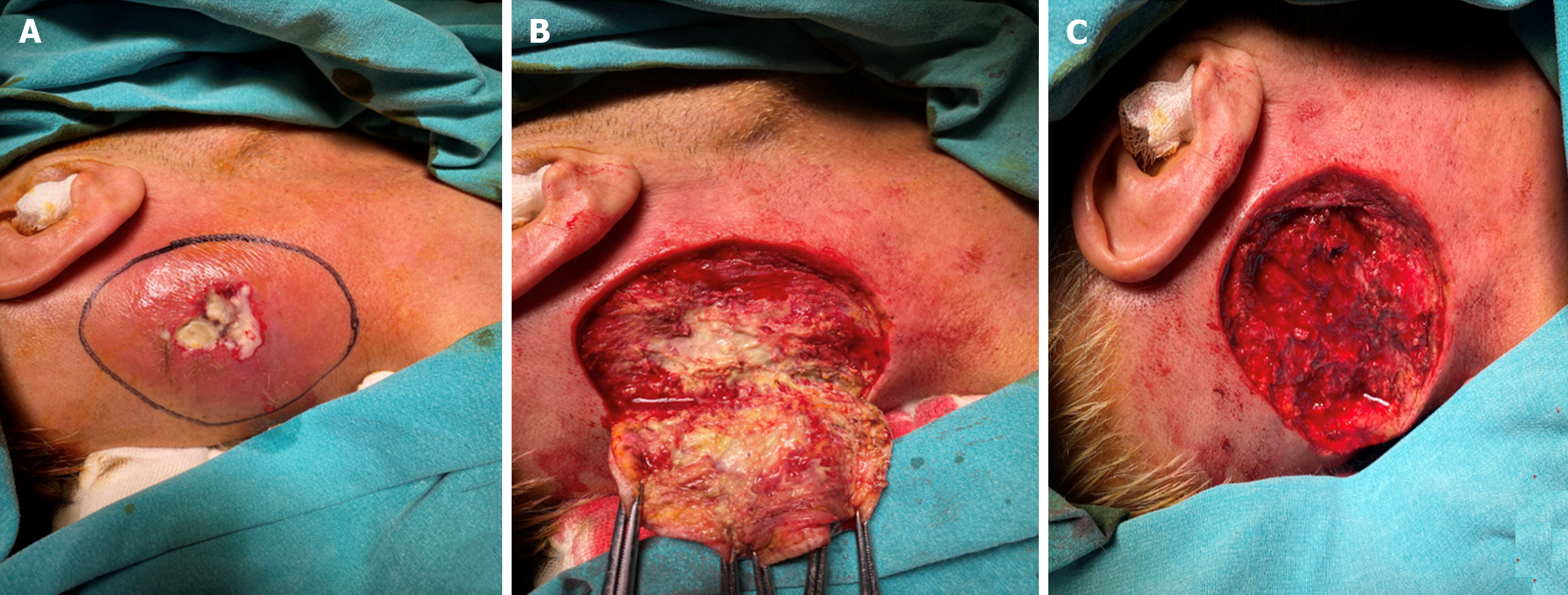
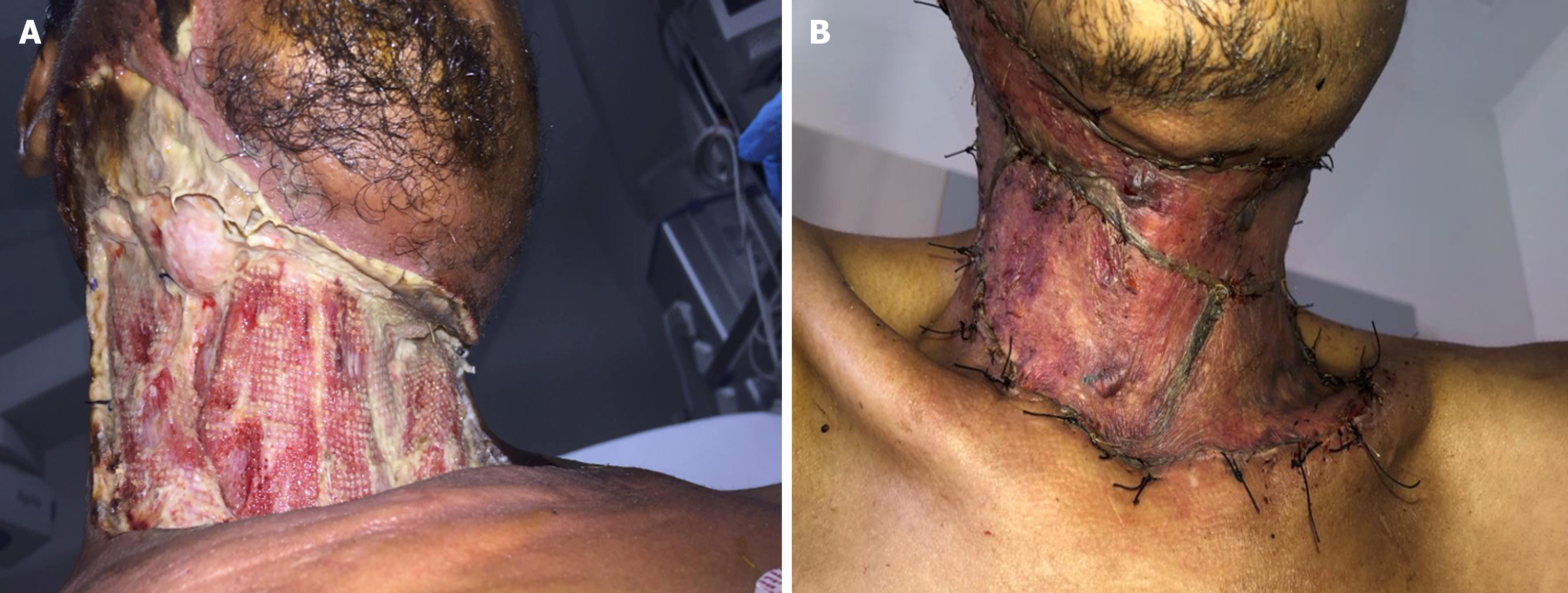
Figure 4 Necrotising fasciitis in posterocervikal region.
A: Preoperative image; B: Intraoperative image; C: Postoperative image.
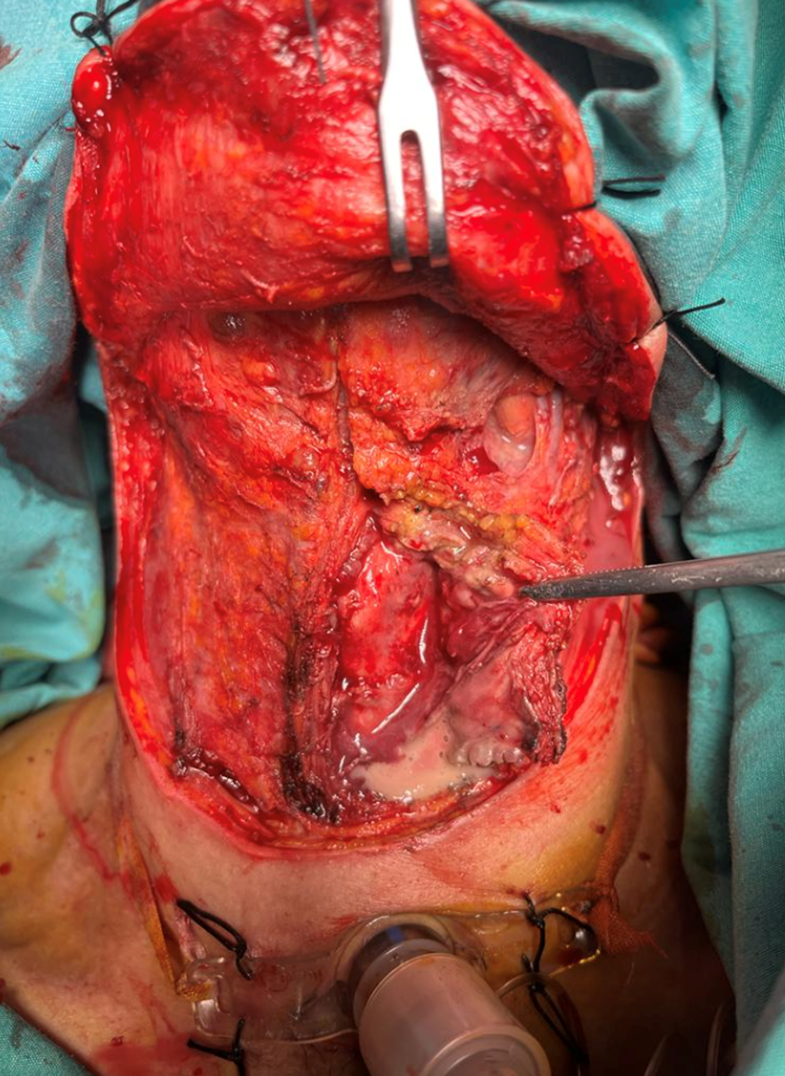
Figure 5
Surgical debridment and tracheotomy due to necrotising fasciitis.
Figure 6 Necrotising fasciitis patient, skin grefting after surgical debridment.
A: After surgical debridment; B: After skin grefting.
- Citation: Bal KK, Aslan C, Gür H, Bal ST, Ustun RO, Unal M. Deep neck infections mortal complications: Intrathoracic complications and necrotising fasciitis. World J Clin Cases 2024; 12(30): 6383-6390
- URL: https://www.wjgnet.com/2307-8960/full/v12/i30/6383.htm
- DOI: https://dx.doi.org/10.12998/wjcc.v12.i30.6383


