Copyright
©The Author(s) 2024.
World J Clin Cases. Oct 16, 2024; 12(29): 6314-6319
Published online Oct 16, 2024. doi: 10.12998/wjcc.v12.i29.6314
Published online Oct 16, 2024. doi: 10.12998/wjcc.v12.i29.6314
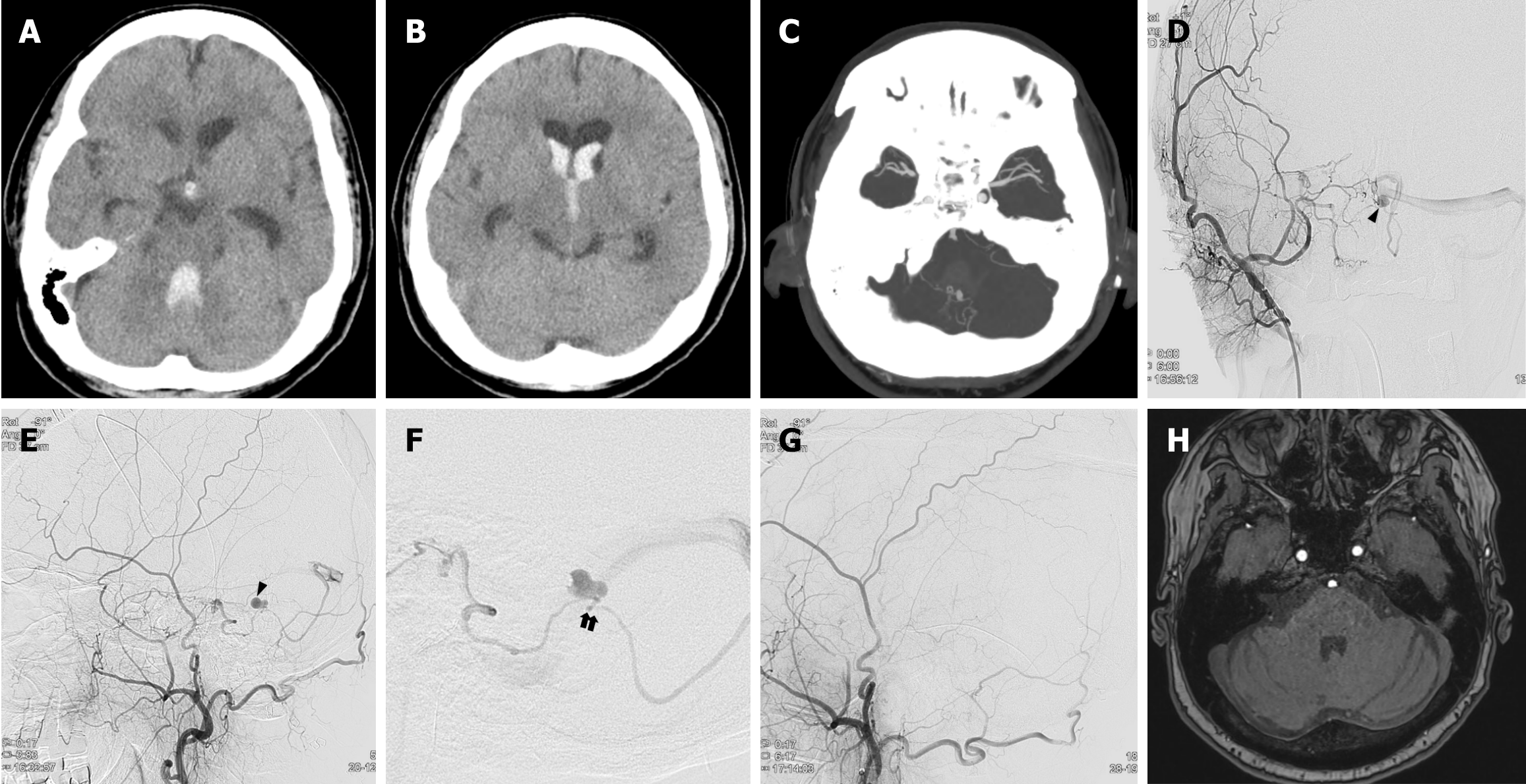
Figure 1 Images of case 1.
A and B: Axial computed tomography (CT) shows acute intraventricular hemorrhage in all ventricles; C: There is an aneurysmal lesion within the 4th ventricle hemorrhage on CT angiography. Right antero-posterior and lateral external carotid artery angiogram showing the dural arteriovenous fistula with venous aneurysm (black arrowhead) fed by petrosal branch of the right middle meningeal artery and drained to left transverse sinus; D and E: There is no evidence of venous ectasia or obstruction in the left transverse sinus; F: Note that the draining vein immediately distal to the aneurysm shows a significant angled appearance (black arrows); G: Transarterial embolization with histoacryl (mixed with lipiodol) was performed and final angiogram shows complete obliteration of the fistula; H: Magnetic resonance angiography, obtained 1 year after embolization, shows no evidence of recurrence with disappearance of venous aneurysm.
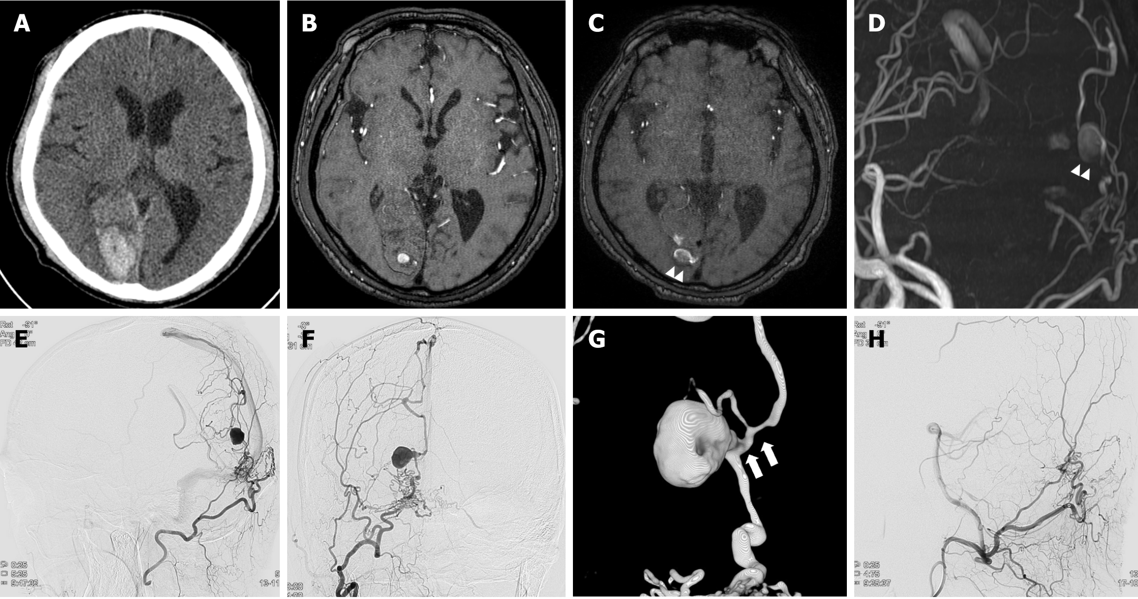
Figure 2 Images of case 2.
A and B: Axial computed tomography and magnetic resonance angiography (MRA), 18 months before admission, shows intracerebral hemorrhage in the right occipital lobe, and there is an aneurysmal lesion within the hemorrhage; C and D: At admission, MRA shows slightly enlarged previous aneurysmal lesion, and this lesion was supplied by a right occipital artery with dilated internal cerebral vein suggesting dural arteriovenous fistula (dAVF) (white arrowheads); E and F: Right antero-posterior and lateral external carotid artery angiogram shows the dAVF with venous aneurysm fed by the right occipital artery and drained to internal cerebral vein and superior sagittal sinus. There is no definite evidence of venous ectasia or obstruction in the venous outlet; G: A rotational 3-dimensional imaging demonstrates a significant curve and stenosis immediately distal to the aneurysm (white arrows); H: Transarterial embolization with histoacryl (mixed with lipiodol) was performed, and the final angiogram showed complete obliteration of the fistula.
- Citation: Kim YS, Yoon W, Baek BH, Kim SK, Joo SP, Kim TS. Ruptured venous aneurysm associated with a dural arteriovenous fistula: Two case reports. World J Clin Cases 2024; 12(29): 6314-6319
- URL: https://www.wjgnet.com/2307-8960/full/v12/i29/6314.htm
- DOI: https://dx.doi.org/10.12998/wjcc.v12.i29.6314


