Copyright
©The Author(s) 2024.
World J Clin Cases. Oct 16, 2024; 12(29): 6307-6313
Published online Oct 16, 2024. doi: 10.12998/wjcc.v12.i29.6307
Published online Oct 16, 2024. doi: 10.12998/wjcc.v12.i29.6307
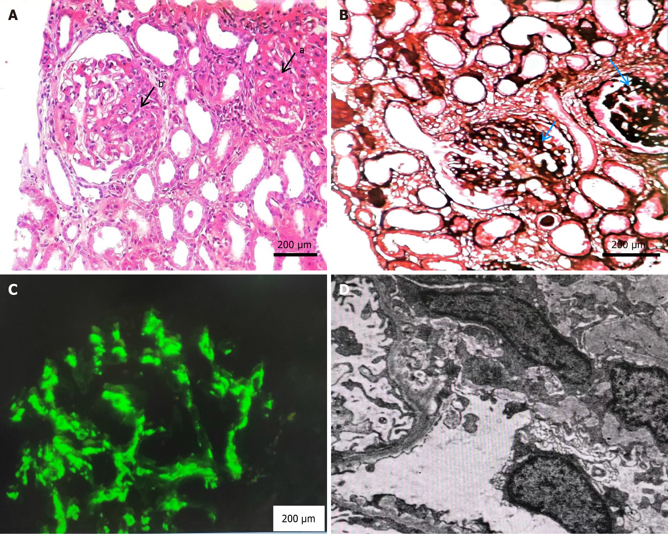
Figure 1 Renal biopsy findings in a patient.
A: Hematoxylin and eosin staining: a-Glomerular ischemic sclerosis; b-Light microscopy image of immunoglobulin A (IgA) (200× magnification); B: Periodic Acid-Schiff Methenamine staining: The basement membrane of glomerular capillaries exhibits extensive vacuolar degeneration (200× magnification); C: Immunofluorescence staining for IgA: IgA deposits in a clustered manner along the mesangial zone (200× magnification); D: Electron microscopy: Deposition of electronic dense substances in the mesangial region (200× magnification).
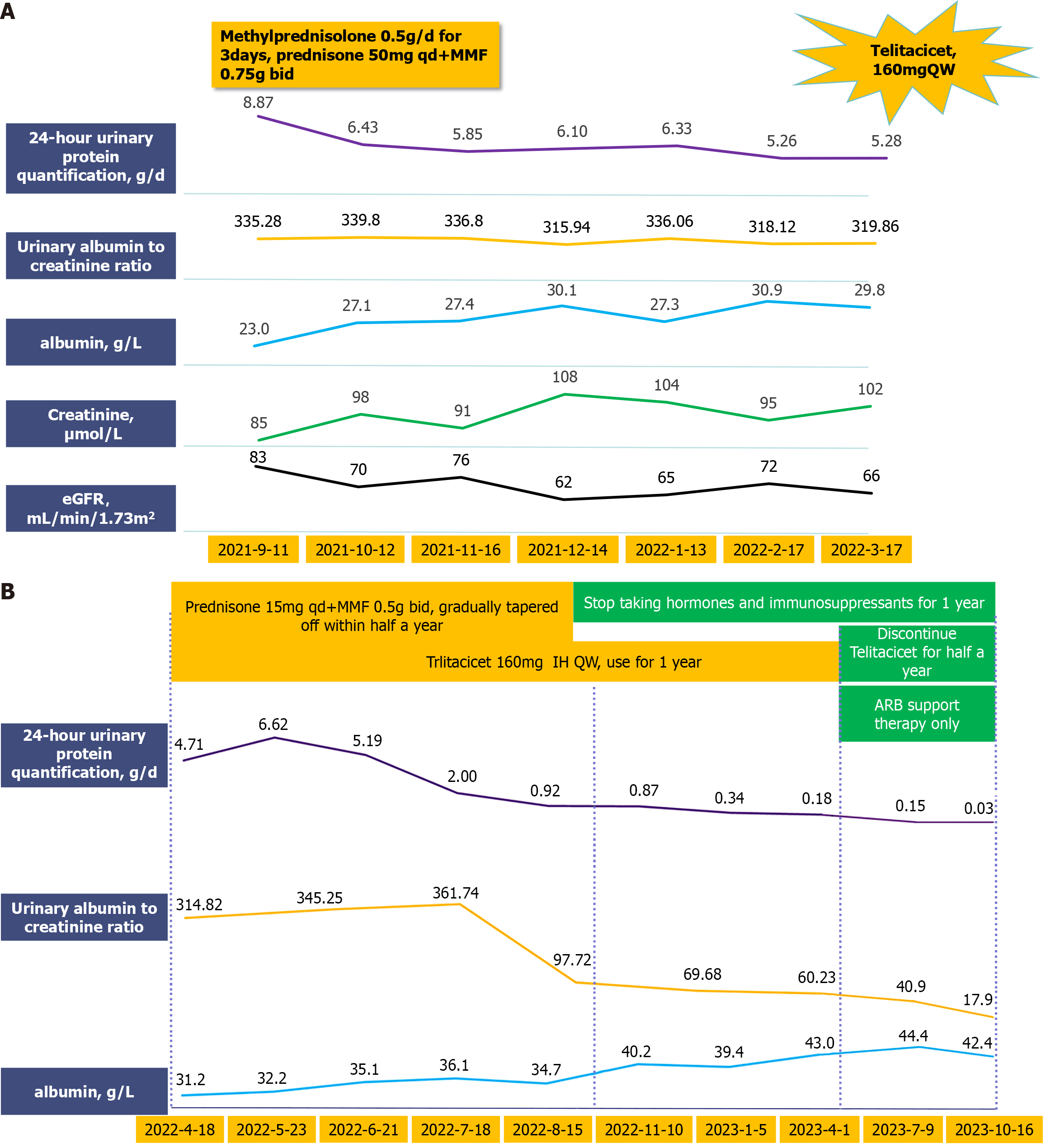
Figure 2 The treatment process received by the patient and changes in indicators.
A: Phase one treatment and results; B: Results and prognosis after adjusting treatment plans.
- Citation: Shen Y, Yuan J, Chen S, Zhang YF, Yin L, Hong Q, Zha Y. Combination treatment with telitacicept, mycophenolate mofetil and glucocorticoids for immunoglobulin A nephropathy: A case report. World J Clin Cases 2024; 12(29): 6307-6313
- URL: https://www.wjgnet.com/2307-8960/full/v12/i29/6307.htm
- DOI: https://dx.doi.org/10.12998/wjcc.v12.i29.6307


