Copyright
©The Author(s) 2024.
World J Clin Cases. Sep 16, 2024; 12(26): 5960-5967
Published online Sep 16, 2024. doi: 10.12998/wjcc.v12.i26.5960
Published online Sep 16, 2024. doi: 10.12998/wjcc.v12.i26.5960
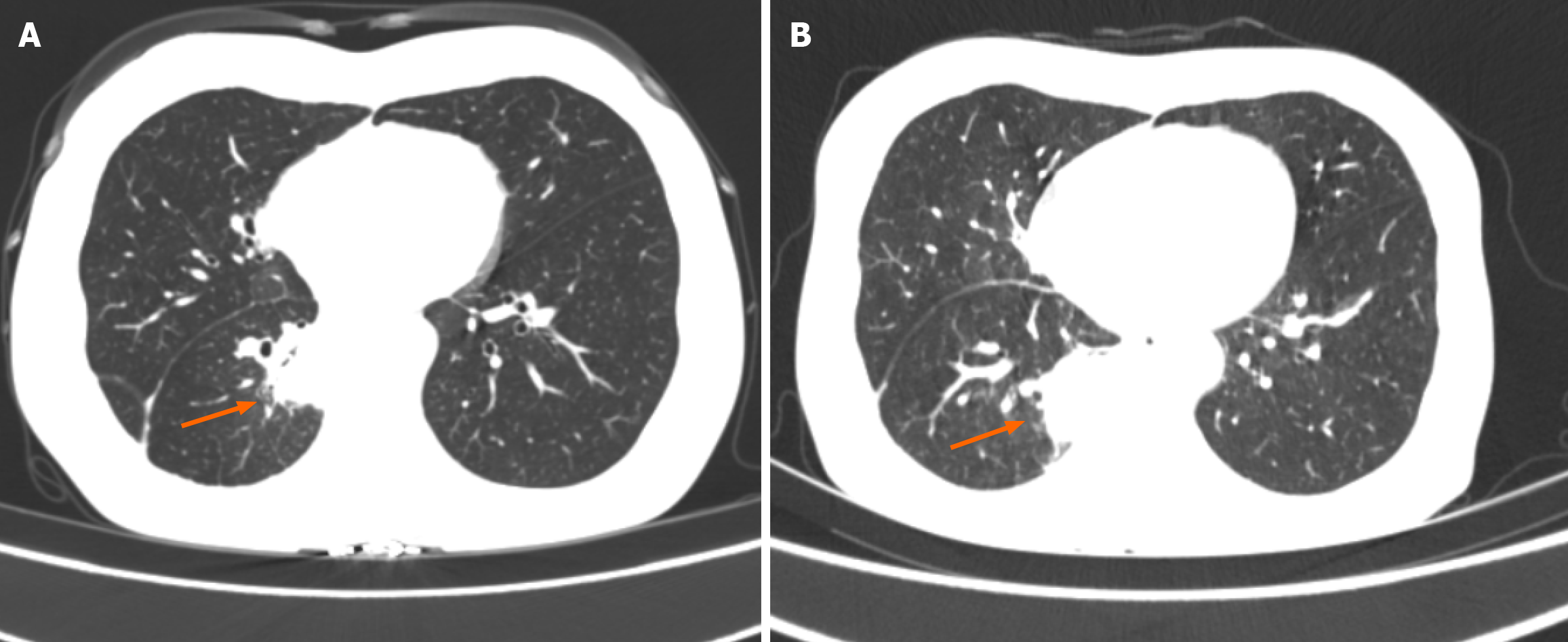
Figure 1 Lung cancer after ablation treatment.
Arrows show the primary lesion. A: Primary site of lung cancer; B: Imaging manifestations after ablative therapy for lung cancer.
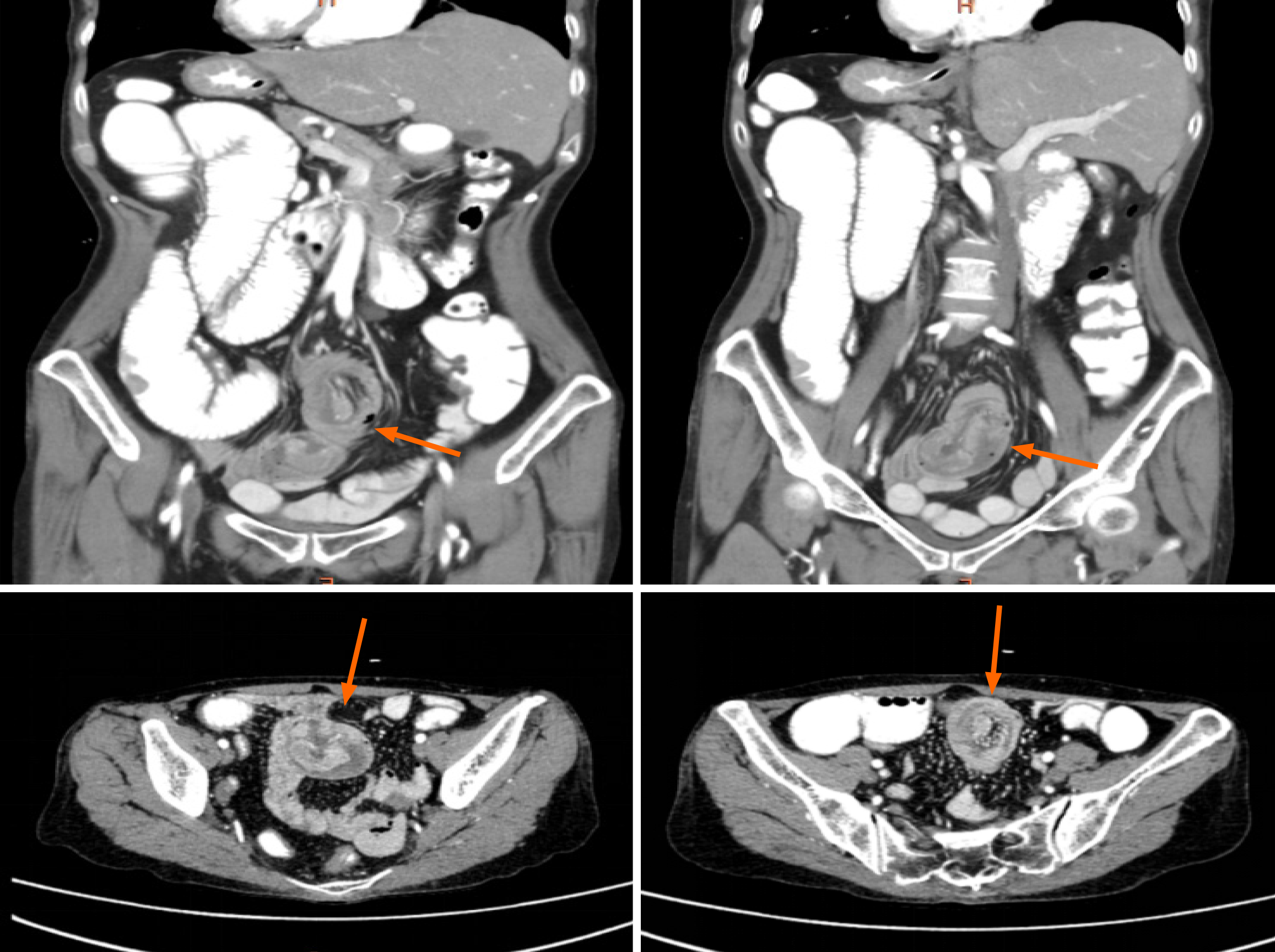
Figure 2 Location of the small bowel intussusception.
Arrows show the location of the small bowel intussusception. A, C, and D: Imaging manifestations of small bowel tumors; B: Location where small bowel obstruction occurred.
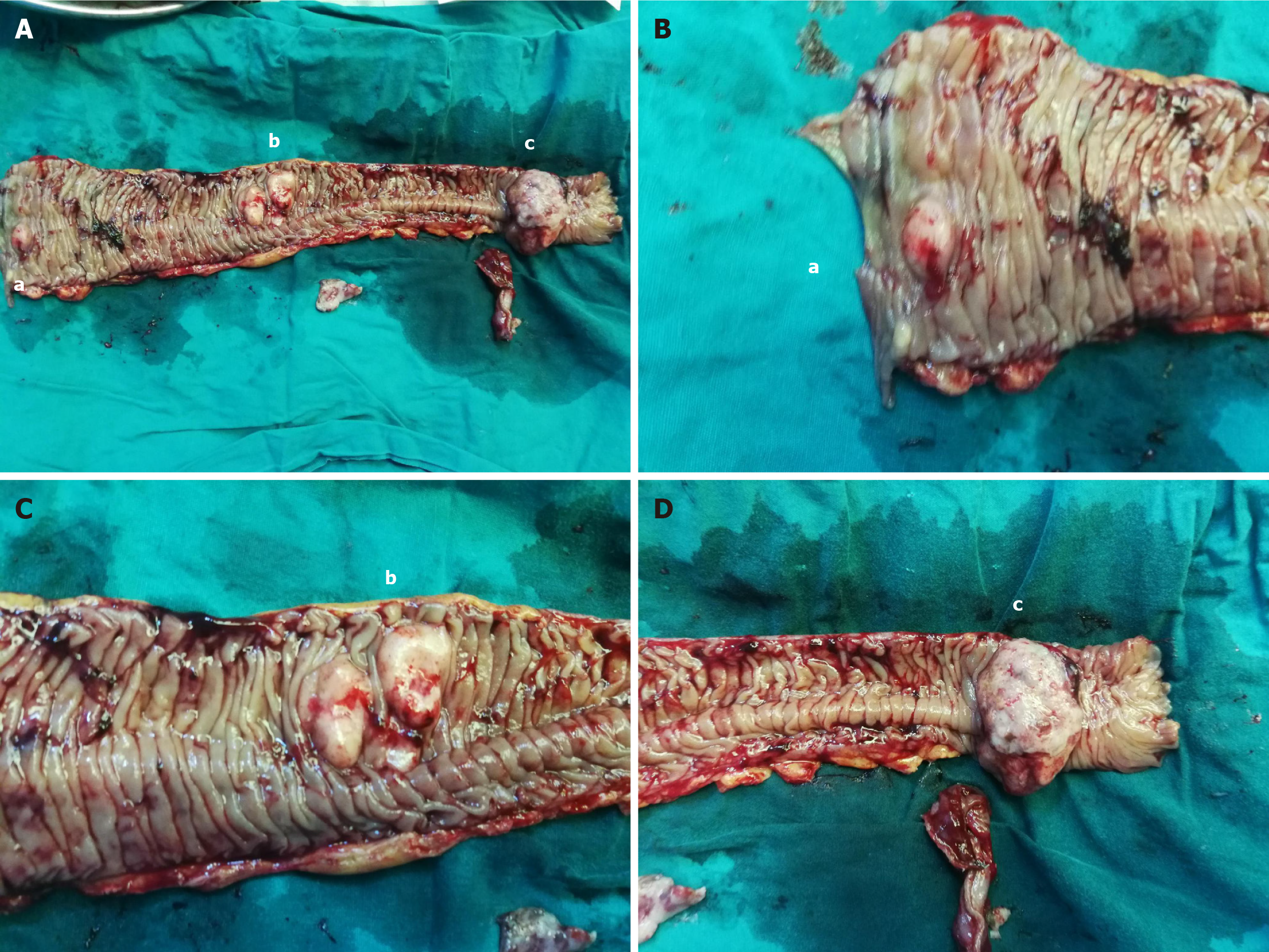
Figure 3 Small bowel tumor.
A: Overall view of the resected small bowel segment that includes three tumors (a, b, and c); B: The smallest small bowel tumor is about 1 cm (a); C: The second largest small bowel tumor (b); D: The largest small bowel tumor was about 6 cm (c).
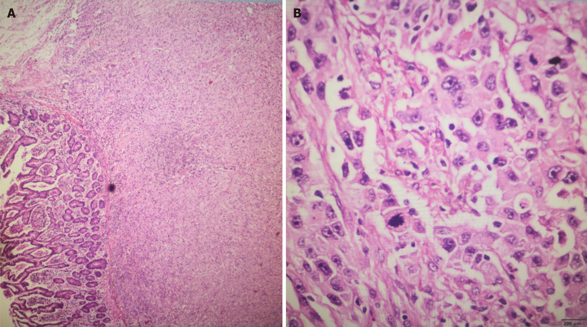
Figure 4 Hematoxylin-eosin staining of tumors of the small intestine.
A: Tumor infiltration to the serosa layer; B: Lung adenocarcinoma metastasis.
Figure 5 Timeline of diagnosis and treatment.
NSCLC: Non-small cell lung cancer.
- Citation: Niu QG, Huang MH, Kong WQ, Yu Y. Stage IV non-small cell lung cancer with multiple metastases to the small intestine leading to intussusception: A case report. World J Clin Cases 2024; 12(26): 5960-5967
- URL: https://www.wjgnet.com/2307-8960/full/v12/i26/5960.htm
- DOI: https://dx.doi.org/10.12998/wjcc.v12.i26.5960


