Copyright
©The Author(s) 2024.
World J Clin Cases. Sep 16, 2024; 12(26): 5908-5921
Published online Sep 16, 2024. doi: 10.12998/wjcc.v12.i26.5908
Published online Sep 16, 2024. doi: 10.12998/wjcc.v12.i26.5908
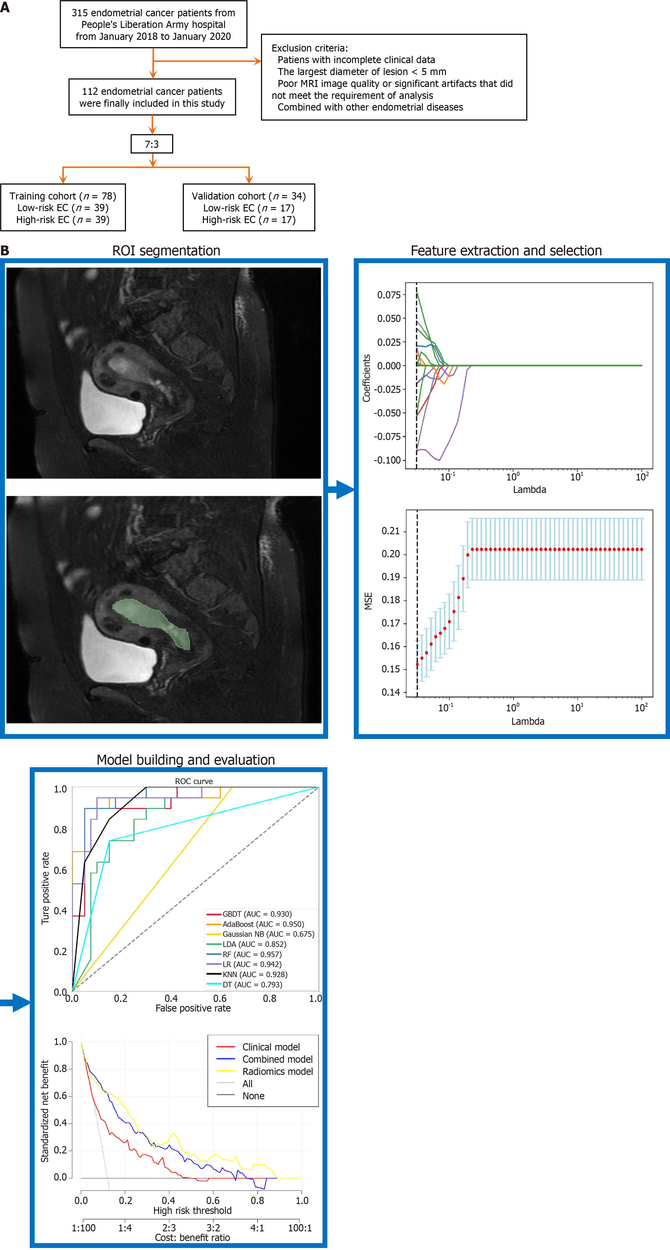
Figure 1 Technology roadmap for this research.
A: Flow chart of patient enrollment; B: Workflow of radiomics analysis process. AUC: Area under the curve; DT: Decision tree; EC: Endometrial cancer; KNN: K-nearest neighbor; LDA: Linear discriminant analysis; LR: Logistic regression; MRI: Magnetic resonance imaging; MSE: Mean-square error; RF: Random forest; ROC: Receiver operating characteristic; ROI: Region of interest.
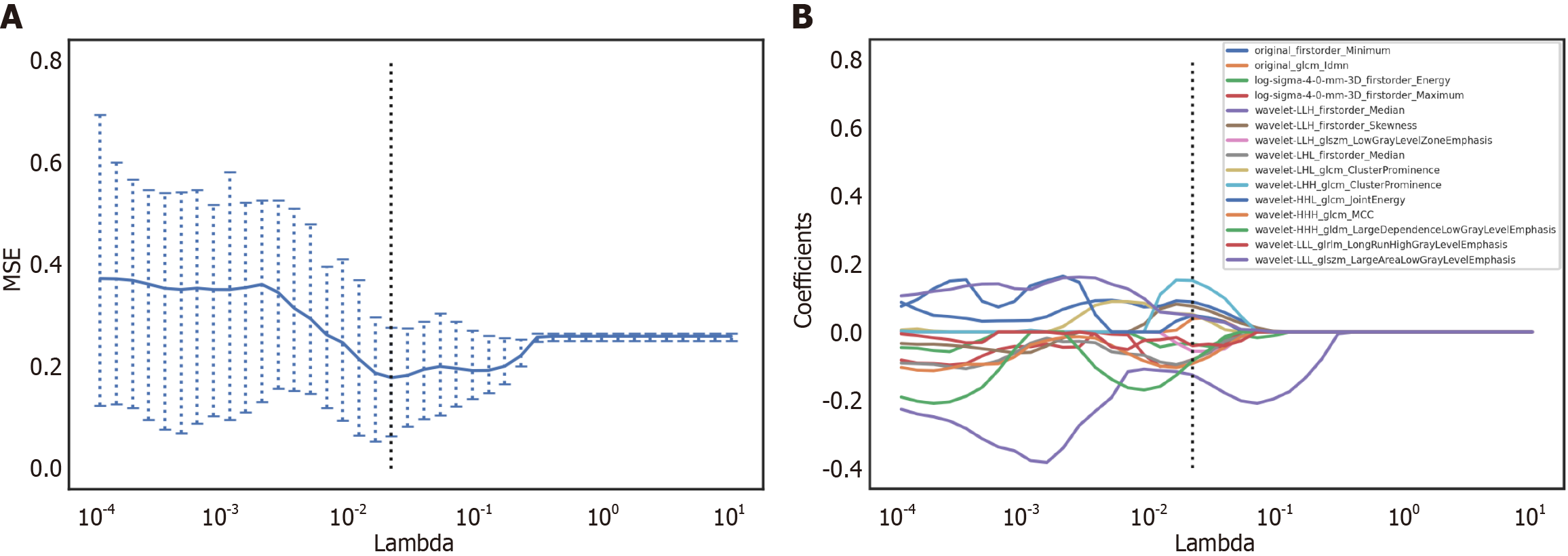
Figure 2 Least absolute shrinkage and selection operator regression model for screening the radiomics characteristics of the training group.
A: Screening of the radiomics features was performed through least absolute shrinkage and selection operator (Lasso) regression. The cross validation for Lasso regression, where the parameter λ was adjusted to find the best function set, is shown. The vertical dotted line on the left panel represents the log(λ) corresponding to the optimal λ; B: Screening of the radiomics features was performed through Lasso regression. The coefficients of texture parameters changed with λ. The vertical line corresponds to the 10 features selected with non-zero Lasso cross-validation coefficients. MSE: Mean-square error.
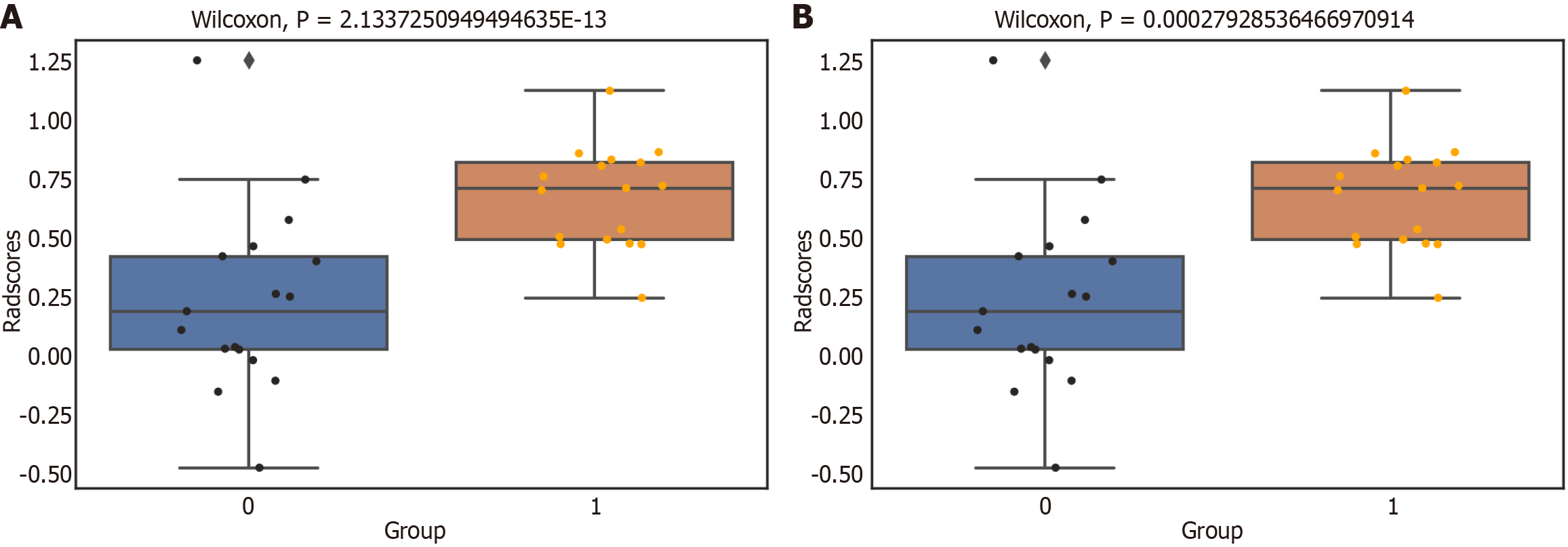
Figure 3 Comparison of low-risk and high-risk endometrial cancer radscores.
A: Training groups; B: Testing groups. 1: Low-risk endometrial cancer; 0: High-risk endometrial cancer.
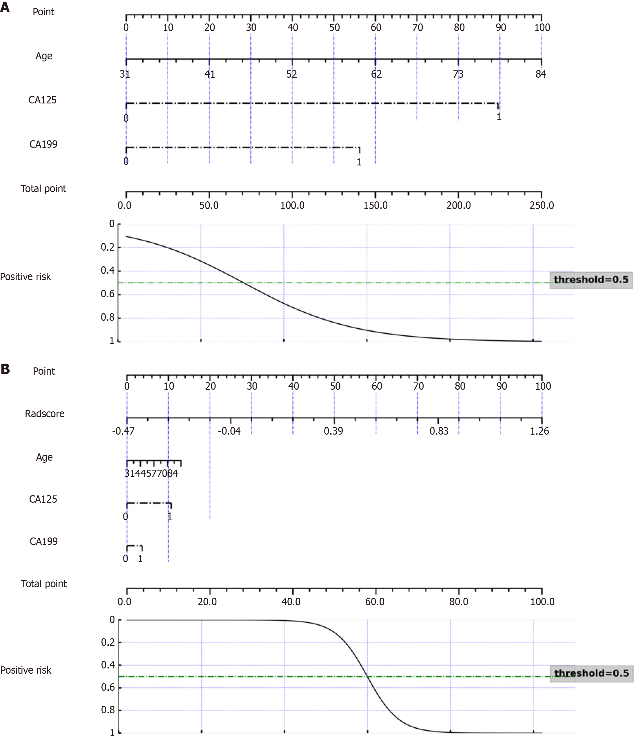
Figure 4 Nomogram to predict endometrial cancer risk.
Cancer antigen (CA) 125 label 1 corresponds to serum levels below 35 U/mL, and label 0 corresponds to serum levels higher than 35 U/mL. CA19-9 label 1 corresponds to serum levels below 27 U/mL, and label 0 corresponds to serum levels higher than 27 U/mL. A: Nomogram developed by clinical predictors; B: Radiomics-clinical combined nomogram.
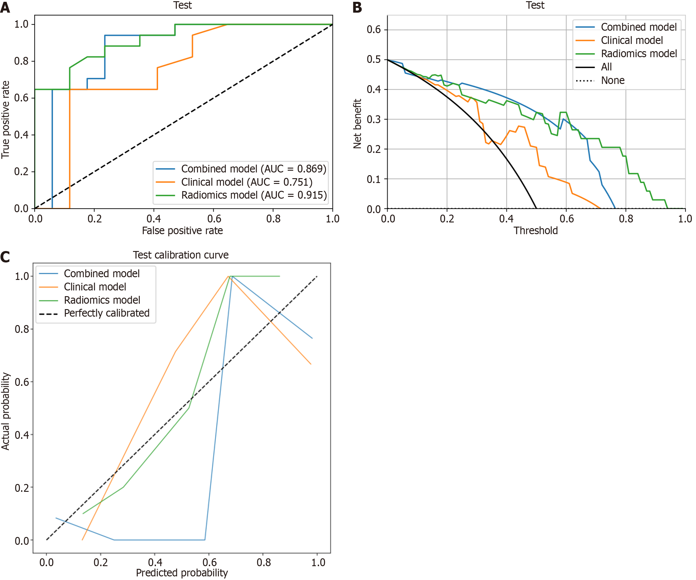
Figure 5 Evaluation of the radiomics model, clinical model, and combined model for predicating endometrial cancer risk grading.
A: Receiver operating characteristic curve; B: Calibration curve; C: Decision curve analysis. AUC: Area under the curve.
- Citation: Wei ZY, Zhang Z, Zhao DL, Zhao WM, Meng YG. Magnetic resonance imaging-based radiomics model for preoperative assessment of risk stratification in endometrial cancer. World J Clin Cases 2024; 12(26): 5908-5921
- URL: https://www.wjgnet.com/2307-8960/full/v12/i26/5908.htm
- DOI: https://dx.doi.org/10.12998/wjcc.v12.i26.5908


