©The Author(s) 2024.
World J Clin Cases. Sep 6, 2024; 12(25): 5821-5831
Published online Sep 6, 2024. doi: 10.12998/wjcc.v12.i25.5821
Published online Sep 6, 2024. doi: 10.12998/wjcc.v12.i25.5821
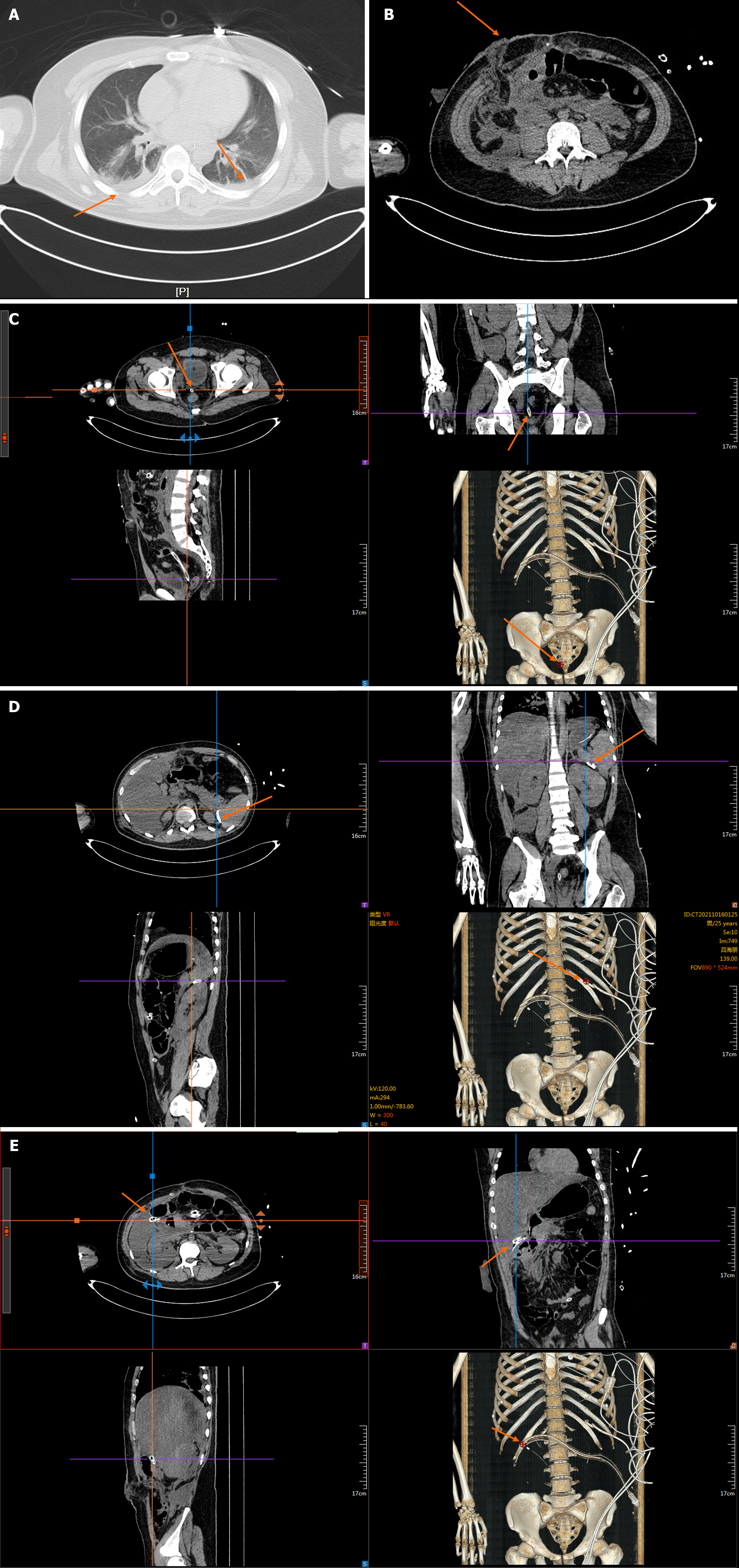
Figure 1 Chest and abdominal computed tomography scans of the patient on admission.
A-E: The patient had pleural effusion (A), a colostomy (B), and abdominal and pelvic drainage tubes (C-E).
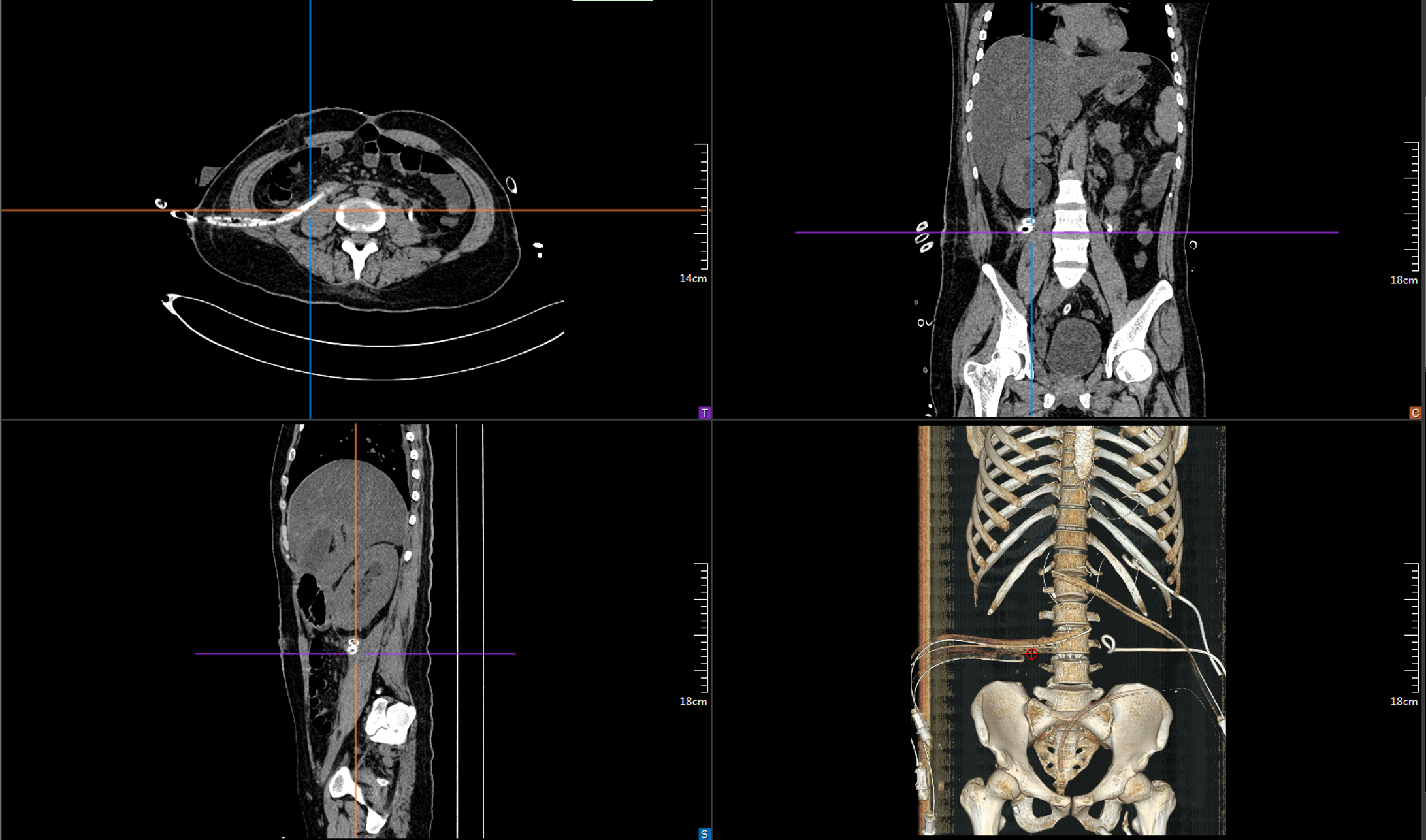
Figure 2 Multiple percutaneous puncture drains were inserted following admission.
A and B: We performed aspiration drainage in the left retroperitoneal (A) and the right retroperitoneal (B) respectively.
Figure 3 Drains placed during surgery.
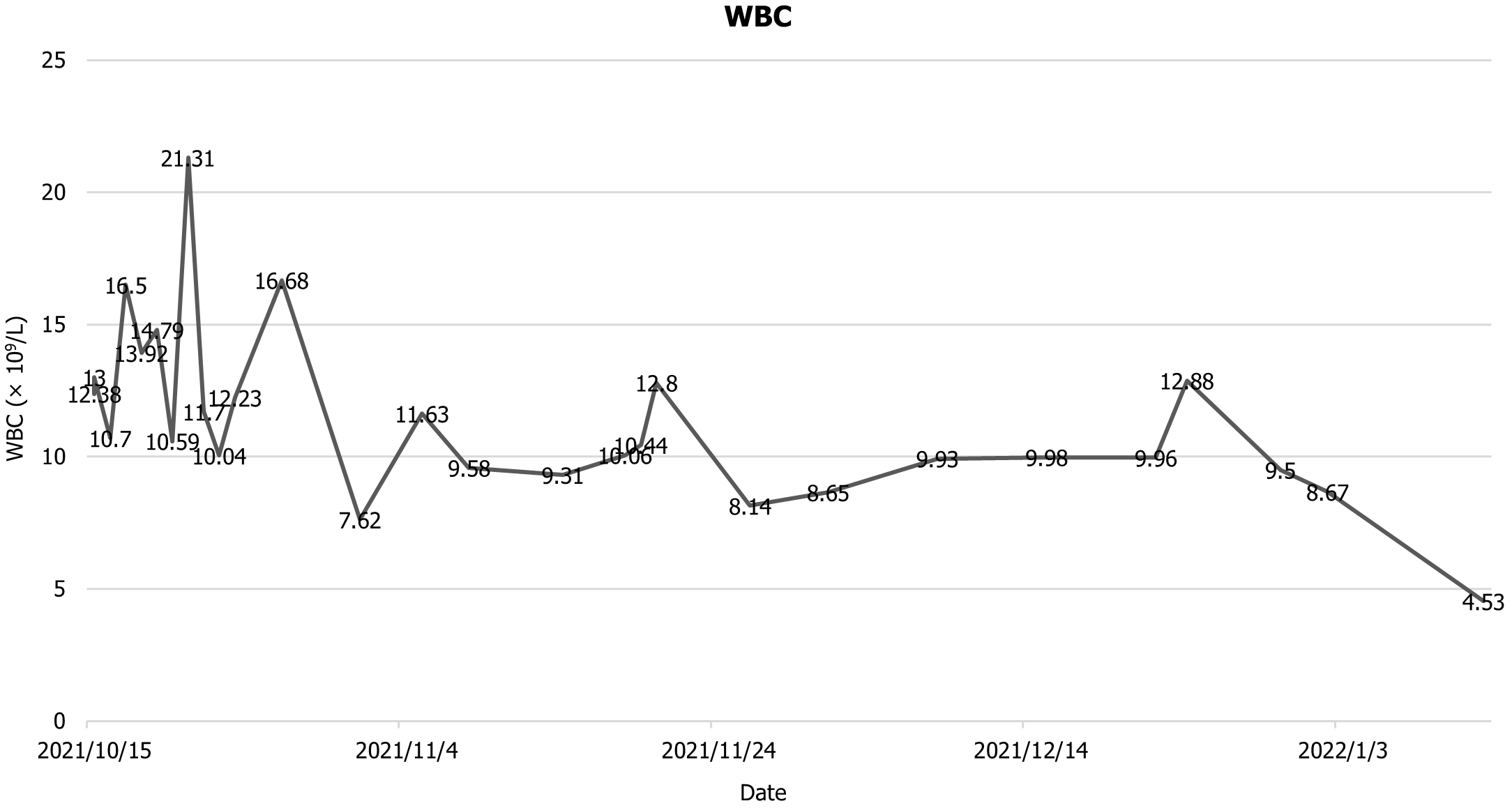
Figure 4 Patient’s white blood cell count during hospitalisation.
WBC: White blood cell.
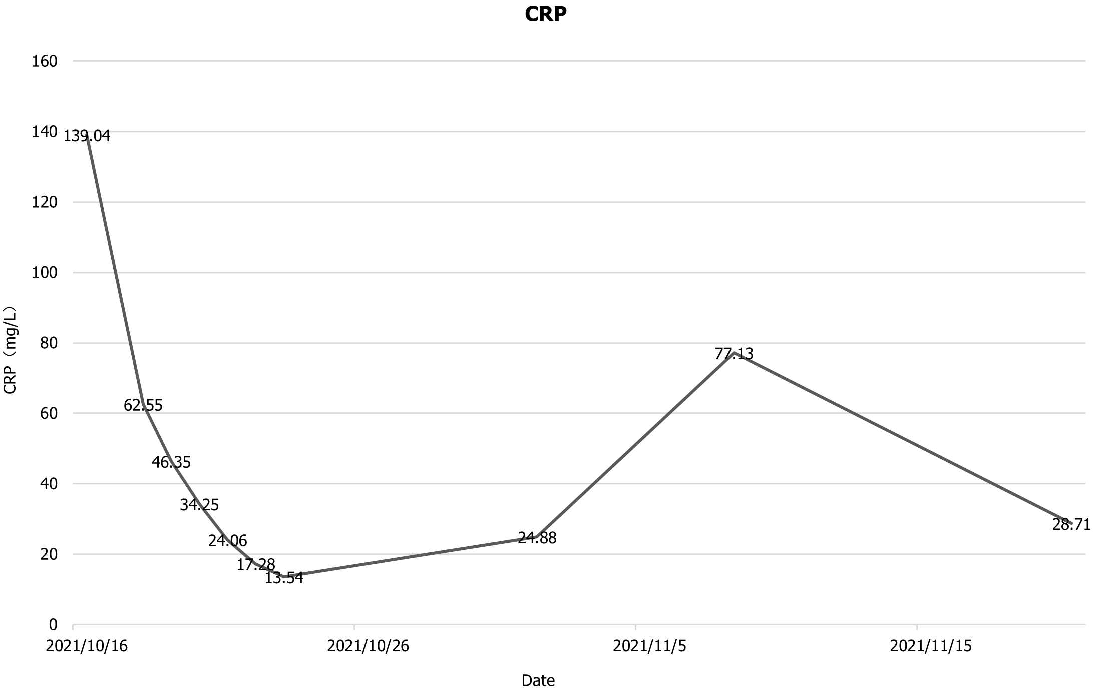
Figure 5 Patient’s C-reactive protein count level during hospitalisation.
CRP: C-reactive protein count.
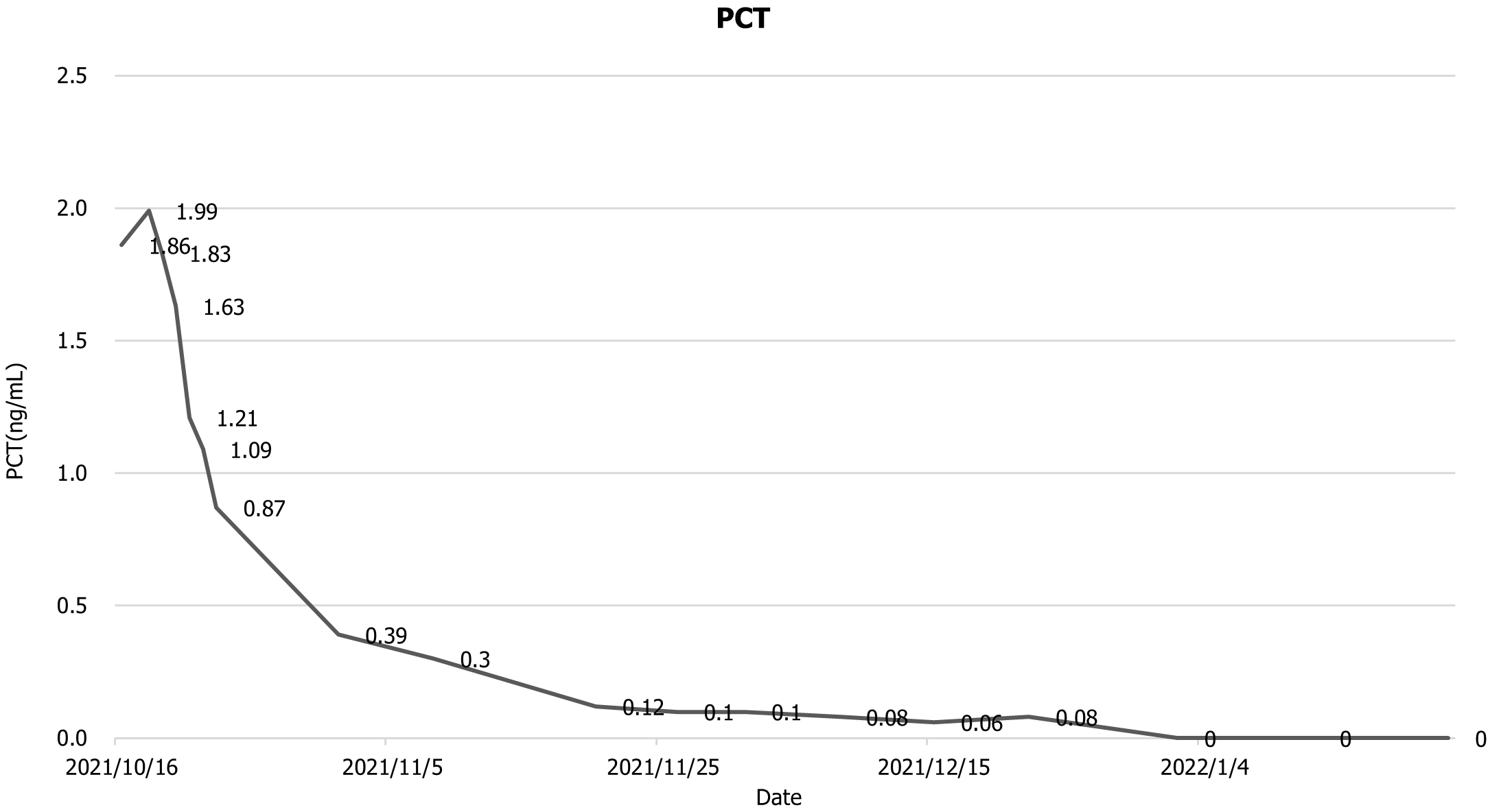
Figure 6 Patient’s procalcitonin count during hospitalisation.
PCT: Procalcitonin.
Figure 7 Flow chart of our team's surgical procedure.
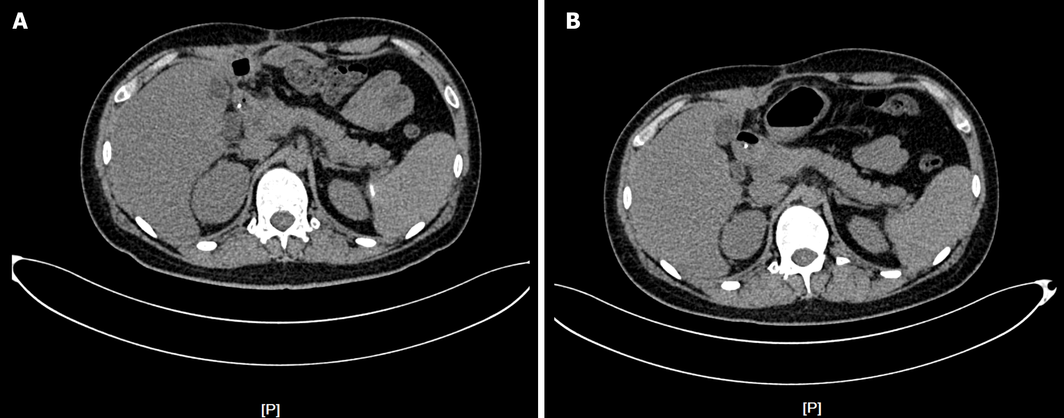
Figure 8 Abdominal computed tomography scan 3 months and 4 months after abdominal trauma.
A: 3 months; B: 4 months.
- Citation: Zhang Y, Cui YF. Severe acute pancreatitis complicated with intra-abdominal infection secondary to trauma: A case report. World J Clin Cases 2024; 12(25): 5821-5831
- URL: https://www.wjgnet.com/2307-8960/full/v12/i25/5821.htm
- DOI: https://dx.doi.org/10.12998/wjcc.v12.i25.5821


