©The Author(s) 2024.
World J Clin Cases. Sep 6, 2024; 12(25): 5814-5820
Published online Sep 6, 2024. doi: 10.12998/wjcc.v12.i25.5814
Published online Sep 6, 2024. doi: 10.12998/wjcc.v12.i25.5814
Figure 1 Preoperative and postoperative magnetic resonance imaging of patients.
A-C: Preoperative magnetic resonance imaging (MRI) indicated a space-occupying lesion in the right cerebellum; D-F: Postoperative follow-up MRI showed that the lesion had been resected.
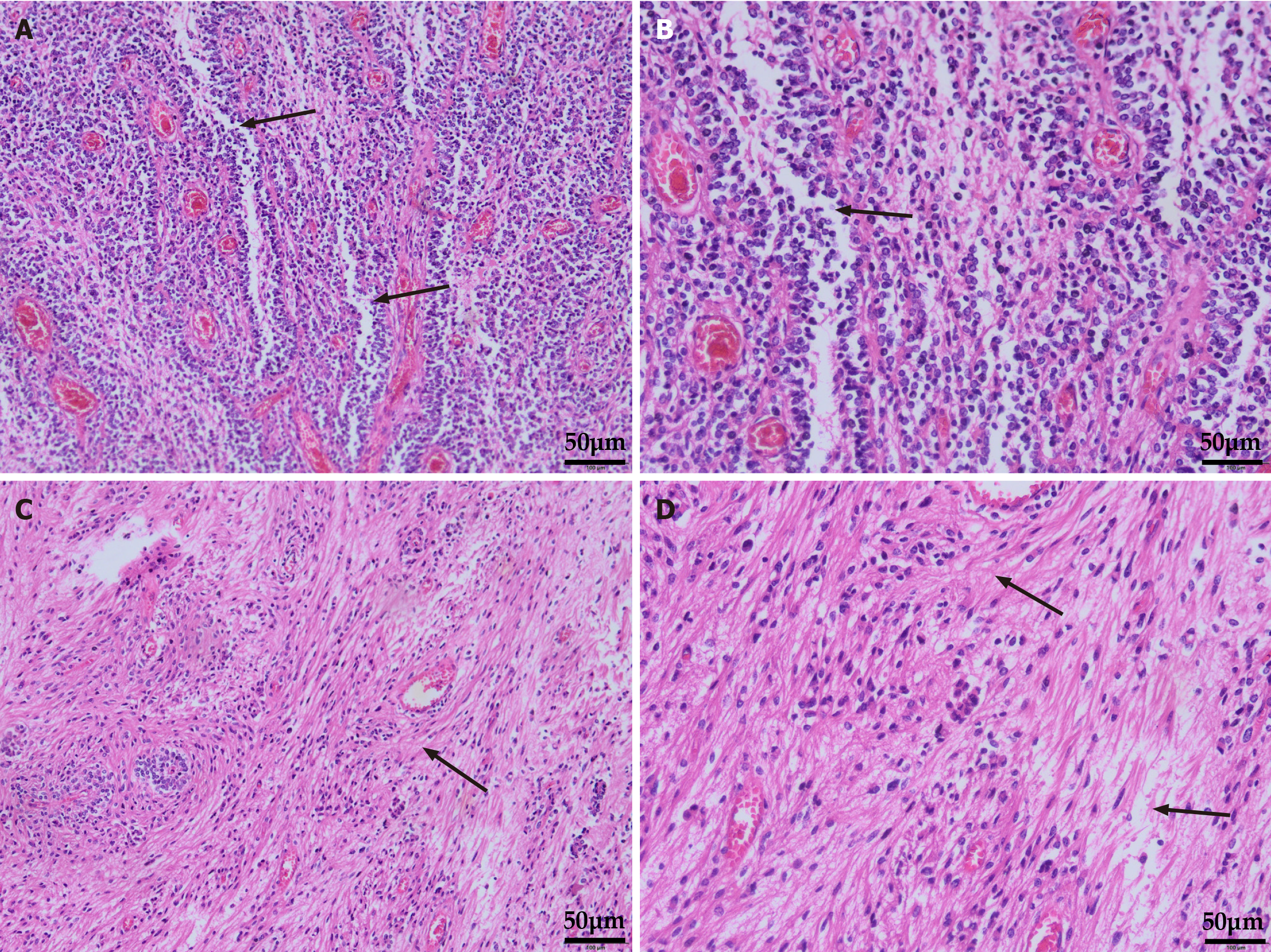
Figure 2 Hematoxylin eosin staining of postoperative tumor tissue sections.
A-B: Ependymal fissure visible; C-D: The arrangement of cells around the blood vessels is blurred, and the cytoplasm gathers around the blood vessels.
Figure 3 Postoperative immunohistochemical analysis of tumor tissue.
GFAP: Glial fibrillary acidic protein; Olig2: Oligodendrocyte transcription factor 2.
- Citation: Yang CG, Xue RF, Yang LX, Jieda XL, Xiang W, Zhou J. Ventricular system-unrelated cerebellar ependymoma: A case report. World J Clin Cases 2024; 12(25): 5814-5820
- URL: https://www.wjgnet.com/2307-8960/full/v12/i25/5814.htm
- DOI: https://dx.doi.org/10.12998/wjcc.v12.i25.5814


