©The Author(s) 2024.
World J Clin Cases. Sep 6, 2024; 12(25): 5798-5804
Published online Sep 6, 2024. doi: 10.12998/wjcc.v12.i25.5798
Published online Sep 6, 2024. doi: 10.12998/wjcc.v12.i25.5798
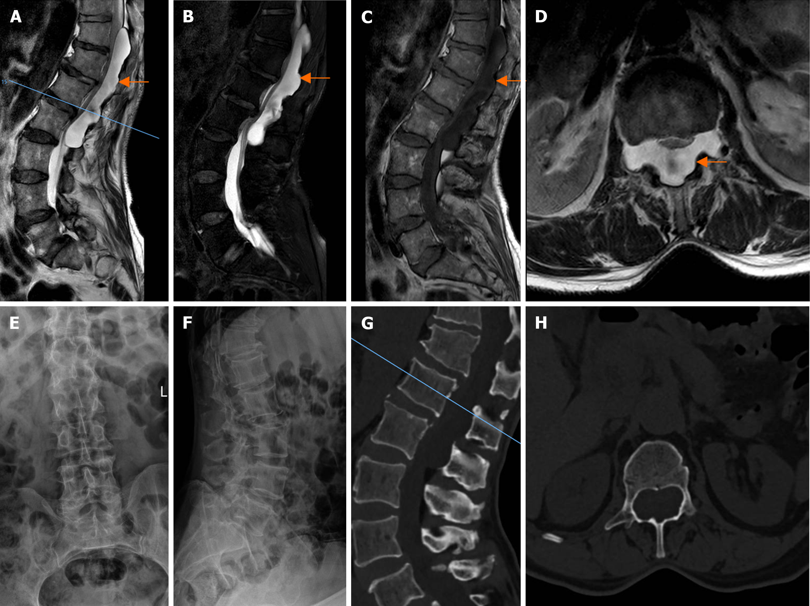
Figure 1 Preoperative imaging data of the patient.
A: Magnetic resonance imaging (MRI) sagittal showed a huge intradural mass in the spinal canal at the level of thoracic 11 lumbar 2; B: High signal in the pressure-lipid sequence; C: Low signal in T1WI; D: MRI axial T2WI showed a high signal in the intradural lesion in the spinal canal; E: Frontal X-ray showed lumbar spine degeneration; F: Lateral X-ray showed instability of T12; G: Computed tomography (CT) reconstruction of sagittal position showed enlarged spinal canal, and the pedicles, plates; H: CT transverse section showed enlargement of the spinal canal with thinning of the pedicle and vertebral plate.
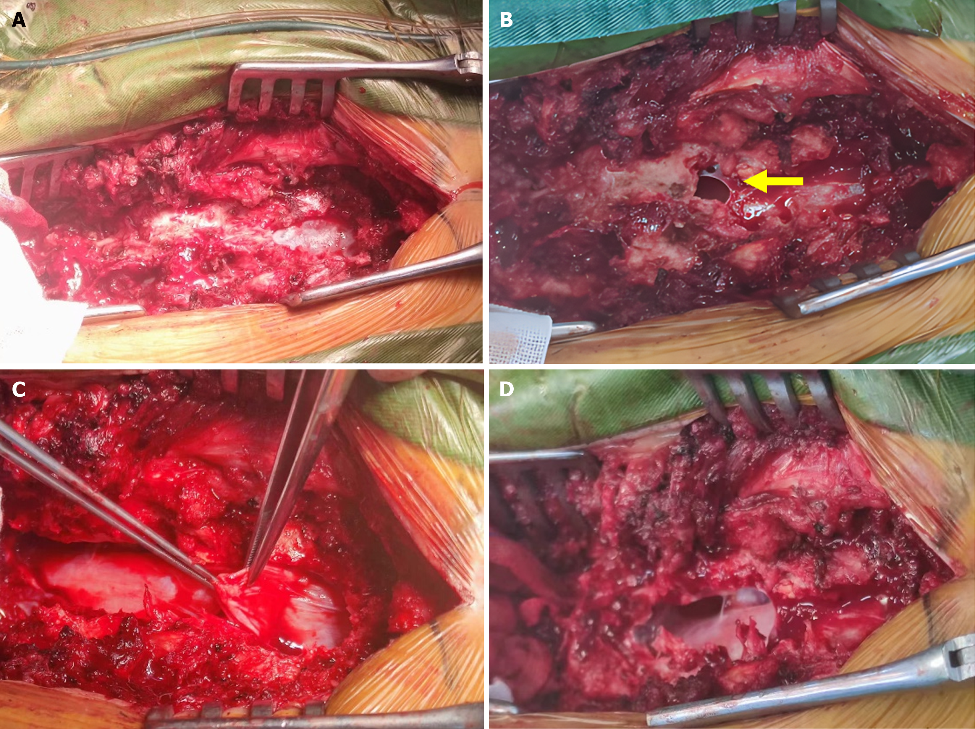
Figure 2 Intraoperative resection process of the cyst.
A: Remove the posterior wall of the spinal canal to fully reveal the head and tail of the cyst as well as both sides; B: The cyst is huge with a thin outer membrane, tearing the wall of the cyst to release its inner clear fluid to reduce the volume of the cyst; C: Carefully peel off the mass, and its outer membrane is seen to be adhered to the dura mater; D: After complete peeling off of the mass, the dura mater is seen to be well pulsed.
Figure 3 Histopathological analysis of the resected specimen.
A: Approximately 70% lymphovascular tissue and 30% vascular tissue at 40 ×; B: Lymphatic venous malformation at 40 ×, manifested by abnormal dilatation, increase, and tortuosity of the vasculature; C: Lymphovascular tissue demonstrated at 100 ×; D: Capillary tissue demonstrated at 100 ×, no abnormal endothelial cell proliferation was seen.
Figure 4 Postoperative imaging.
A: Frontal X-ray radiographs showed internal fixation; B: Lateral X-ray radiographs showed the internal fixation; C: Computed tomography (CT) sagittal view showing internal fixation with pedicle root nail rods; D: CT transverse view of the vertebral pedicle in good position.
- Citation: Sun SF, Wang XH, Yuan YY, Shao YD. Rare giant intradural epidural hemolymphangioma: A case report. World J Clin Cases 2024; 12(25): 5798-5804
- URL: https://www.wjgnet.com/2307-8960/full/v12/i25/5798.htm
- DOI: https://dx.doi.org/10.12998/wjcc.v12.i25.5798


