©The Author(s) 2024.
World J Clin Cases. Sep 6, 2024; 12(25): 5761-5768
Published online Sep 6, 2024. doi: 10.12998/wjcc.v12.i25.5761
Published online Sep 6, 2024. doi: 10.12998/wjcc.v12.i25.5761
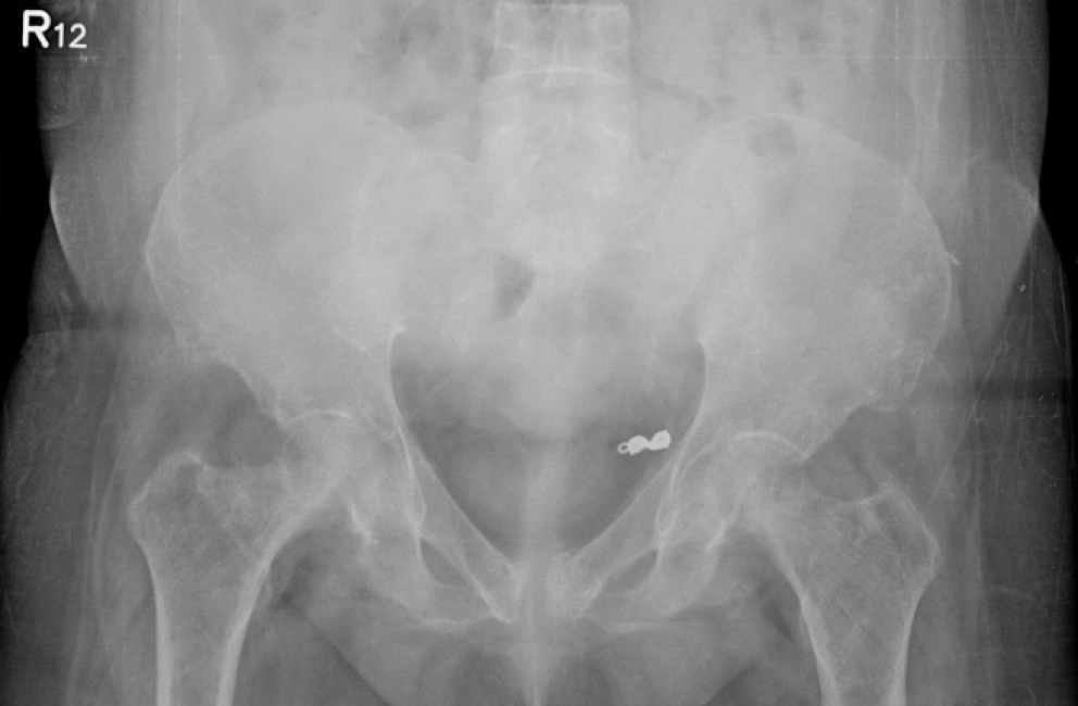
Figure 1 Anteroposterior view of the initial hip X-ray after the fall.
It shows no obvious right femoral neck fracture. Note that the cortical thickness of diaphysis was similar bilaterally.
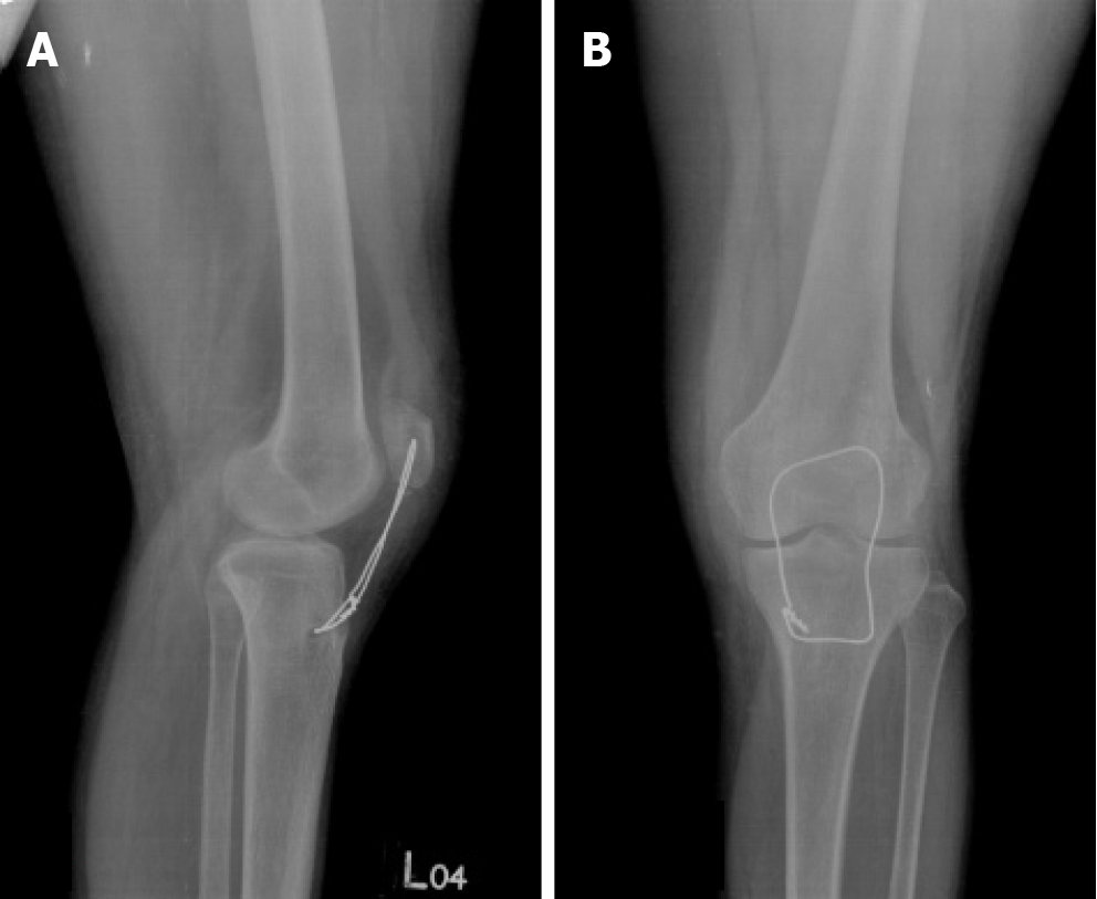
Figure 2 Patellar avulsion fracture status post open reduction internal fixation with cerclage wire.
A: Lateral view; B: Anteroposterior view.
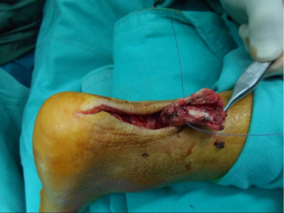
Figure 3 Intraoperative view of the right achilles tendon rupture.
Figure 4 A hip X-ray 1 year after the fall.
It showed a neglected (blue arrow) right femoral neck fracture and marked cortical thinning of the right femoral diaphysis compared with the left (orange arrow) due to stress-shielding.
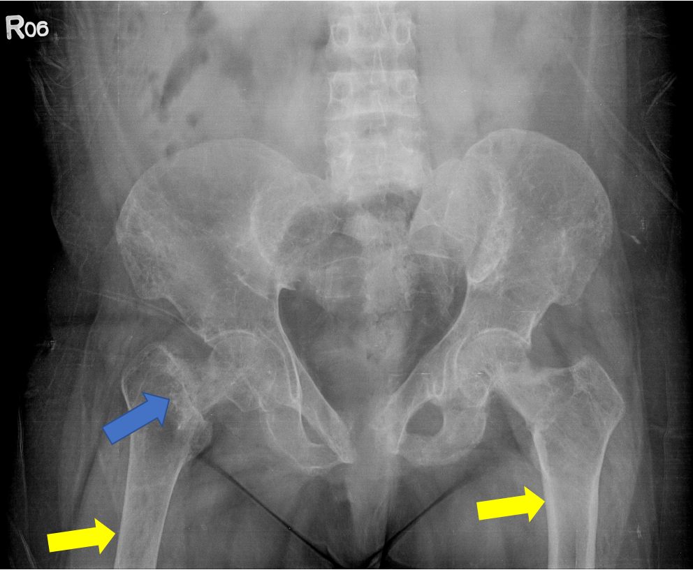
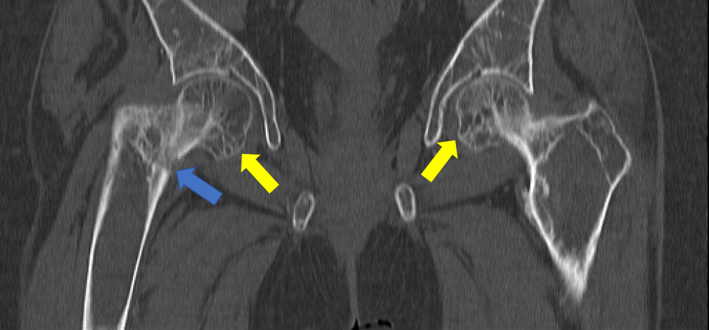
Figure 5 Computed tomography of the hip 1 year after the fall.
It showed a right femur neck fracture (blue arrow) with osteitis fibrosa cystica in the bilateral femoral heads (yellow arrows).
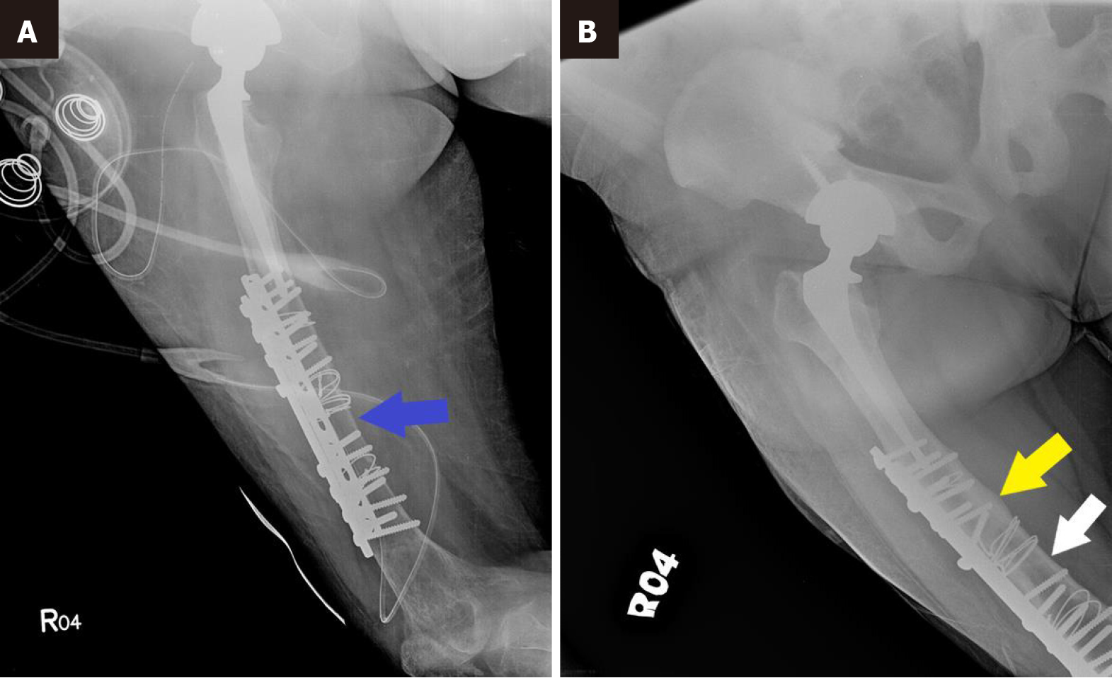
Figure 6 Hip X-rays.
A: Hip X-ray shows thin cortical bone (blue arrow) at the time the fracture occurred; B: Hip X-ray 3 years after subtotal parathyroidectomy and 2 years after right hip replacement shows solid union of the fracture (yellow arrow), with restoration of the cortical bone thickness (white arrow).
Figure 7 Bone mineral density 8 months after subtotal parathyroidectomy and 2 months after right total hip replacement.
A: Vertebral body; B: Left femoral neck. BMD: Bone mineral density.
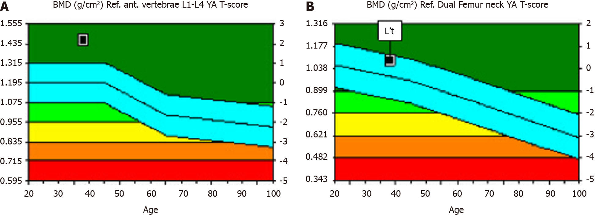
Figure 8 Bone mineral density 3 years after subtotal parathyroidectomy and 2 years after right total hip replacement.
A: Vertebral body; B: Left femoral neck, note that bone mineral density of the lumbar spine (L1-L4) increased by 59%. BMD: Bone mineral density.
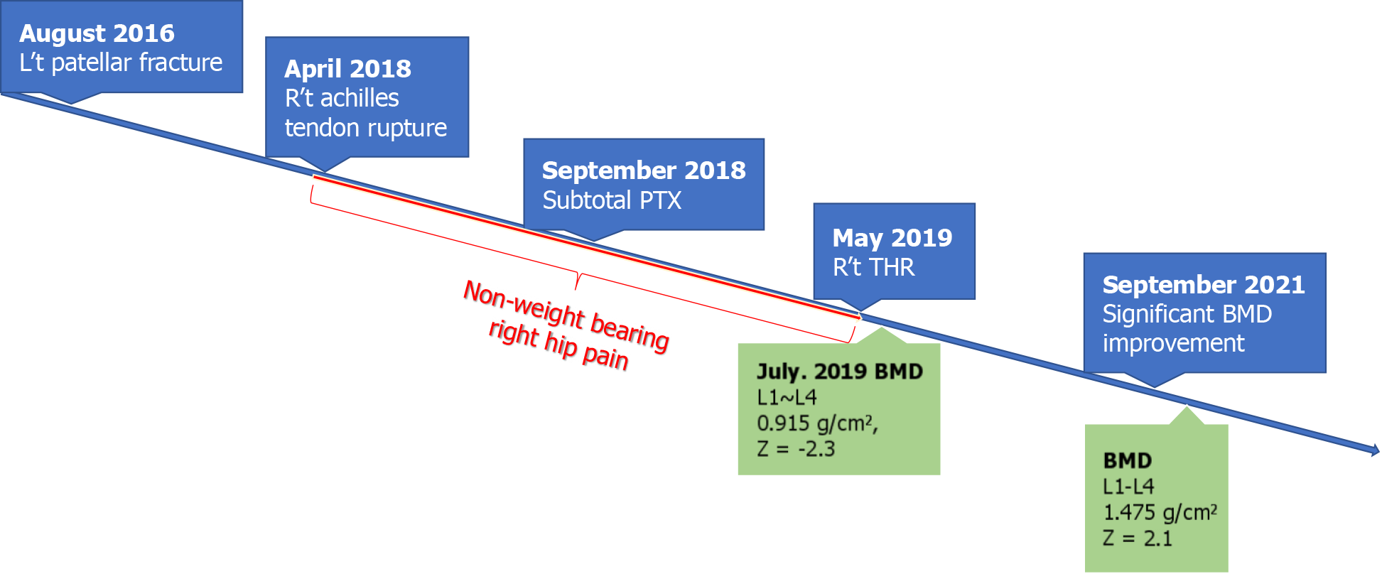
Figure 9 Timeline of the clinical course.
PTX: Parathyroidectomy; THR: Hip replacement ; BMD: Bone mineral density.
- Citation: Lin TC, Lin SW, Yeh KT. Parathyroidectomy restored bone mineral density in a neglected femoral neck fracture with renal osteodystrophy: A case report. World J Clin Cases 2024; 12(25): 5761-5768
- URL: https://www.wjgnet.com/2307-8960/full/v12/i25/5761.htm
- DOI: https://dx.doi.org/10.12998/wjcc.v12.i25.5761


