©The Author(s) 2024.
World J Clin Cases. Jul 16, 2024; 12(20): 4446-4451
Published online Jul 16, 2024. doi: 10.12998/wjcc.v12.i20.4446
Published online Jul 16, 2024. doi: 10.12998/wjcc.v12.i20.4446
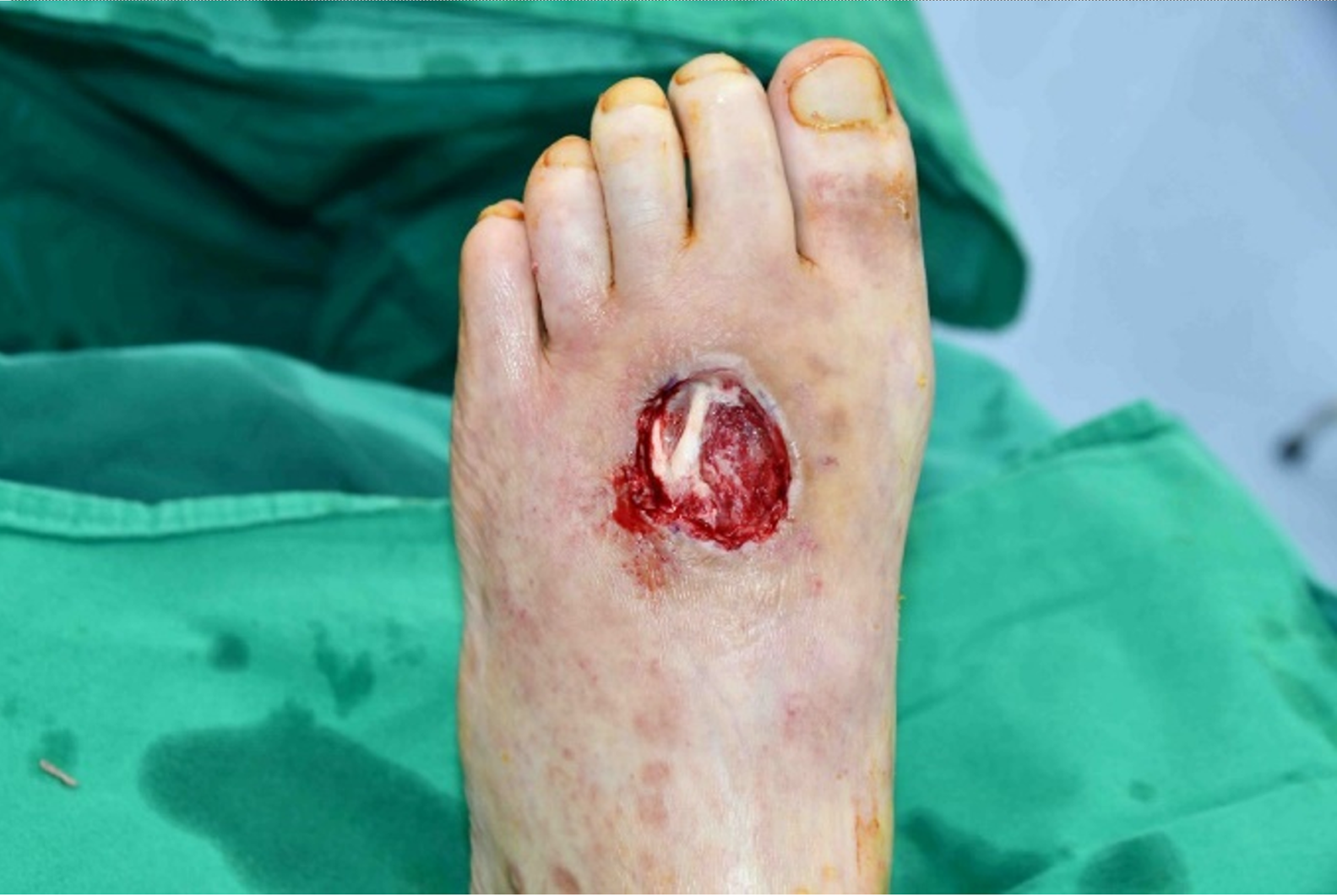
Figure 1 Case 1, pretreatment photograph after initial debridement.
The diabetic foot ulcer measuring 3 cm 3 cm on the dorsum of the left foot with exposure of muscle and tendon (Wagner grade II).
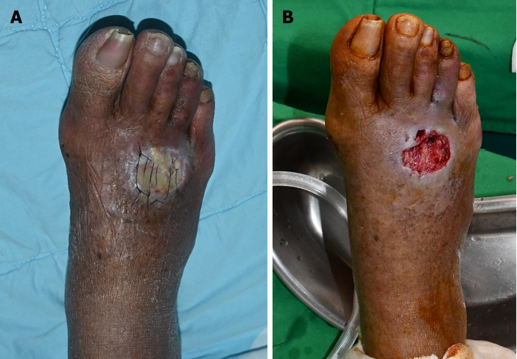
Figure 2 Case 2, pretreatment photograph before and after initial debridement.
The diabetic foot ulcer measuring 2 cm 2.5 cm on the dorsum of the right foot with exposure of muscle (Wagner grade II). A: Pretreatment photograph before initial debridement; B: Pretreatment photograph after initial debridement.
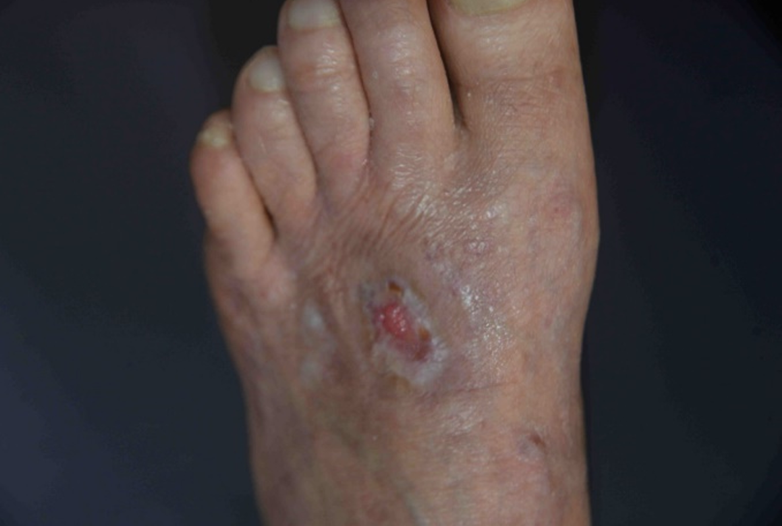
Figure 3 Case 1, post-treatment photograph.
After treatment with polydeoxyribonucleotide injection for 8 wk, the raw surface area was healed and epithelialized without exposure of muscles and tendons.
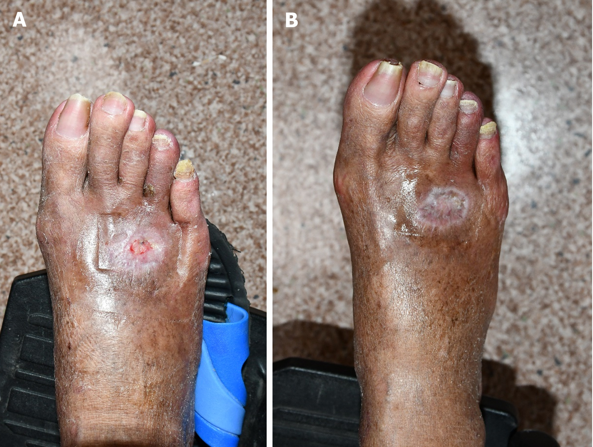
Figure 4 Case 2, post-treatment photograph.
After treatment with polydeoxyribonucleotide injection for 6 wk (left) and 8 wk (right), the raw surface was almost healed by 6 wk and completely healed and epithelialized without exposure of muscles by 8 wk. A: After treatment with polydeoxyribonucleotide injection for 6 wk; B: After treatment with polydeoxyribonucleotide injection for 8 wk.
- Citation: Ha Y, Kim JH, Kim J, Kwon H. Non-surgical treatment of diabetic foot ulcers on the dorsum of the foot with polydeoxyribonucleotide injection: Two case reports. World J Clin Cases 2024; 12(20): 4446-4451
- URL: https://www.wjgnet.com/2307-8960/full/v12/i20/4446.htm
- DOI: https://dx.doi.org/10.12998/wjcc.v12.i20.4446


