©The Author(s) 2024.
World J Clin Cases. Jul 16, 2024; 12(20): 4384-4390
Published online Jul 16, 2024. doi: 10.12998/wjcc.v12.i20.4384
Published online Jul 16, 2024. doi: 10.12998/wjcc.v12.i20.4384
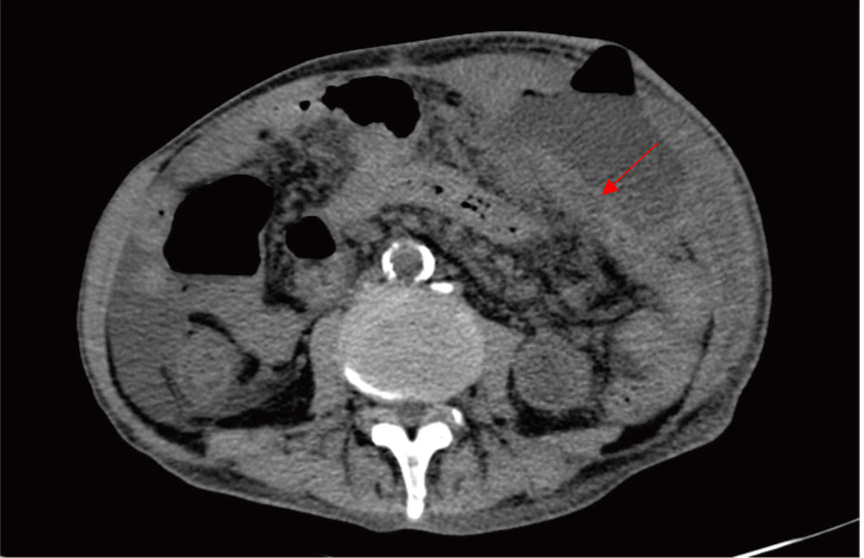
Figure 1 Inflammatory exudate and encapsulated abscess on computed tomography imaging.
Axial computed tomography image showing a ‘track’ (red arrow) crossing from the peritoneal cavity through the anterior abdominal wall.
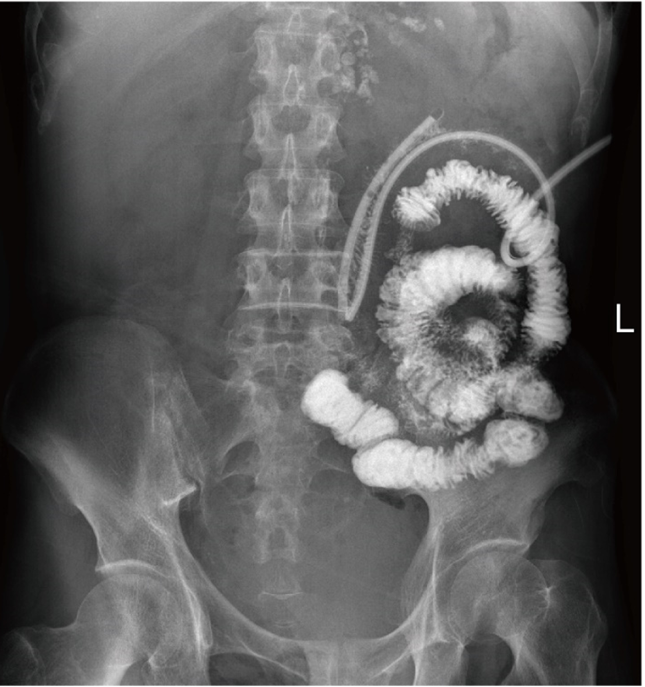
Figure 2 Anatomic position of the fistula confirmed by a fistulogram.
Administration of contrast agent to the intestine suggested a fistula.
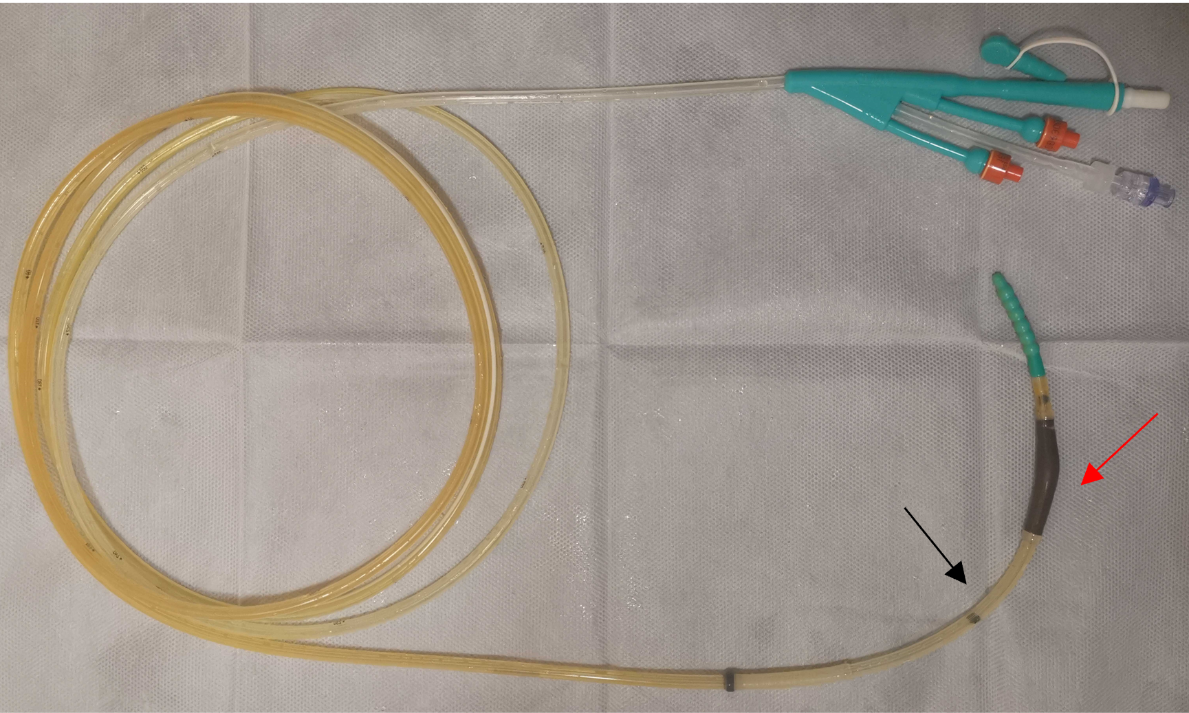
Figure 3 Construction of the intestinal obstruction catheter.
The catheter (model number: 16DBR 3000T0) was a tube with a total length of 3 metres and a diameter of 16Fr and was manufactured by CREAT MEDIC DALIAN International Trading Co., Ltd. It was made of medical silicone rubber. The catheter has an anterior balloon (red arrow) that acts as a counterweight to guide the catheter into the correct position of the intestine, and its suction port (black arrow) can be used for the inflow of nutritional substances.
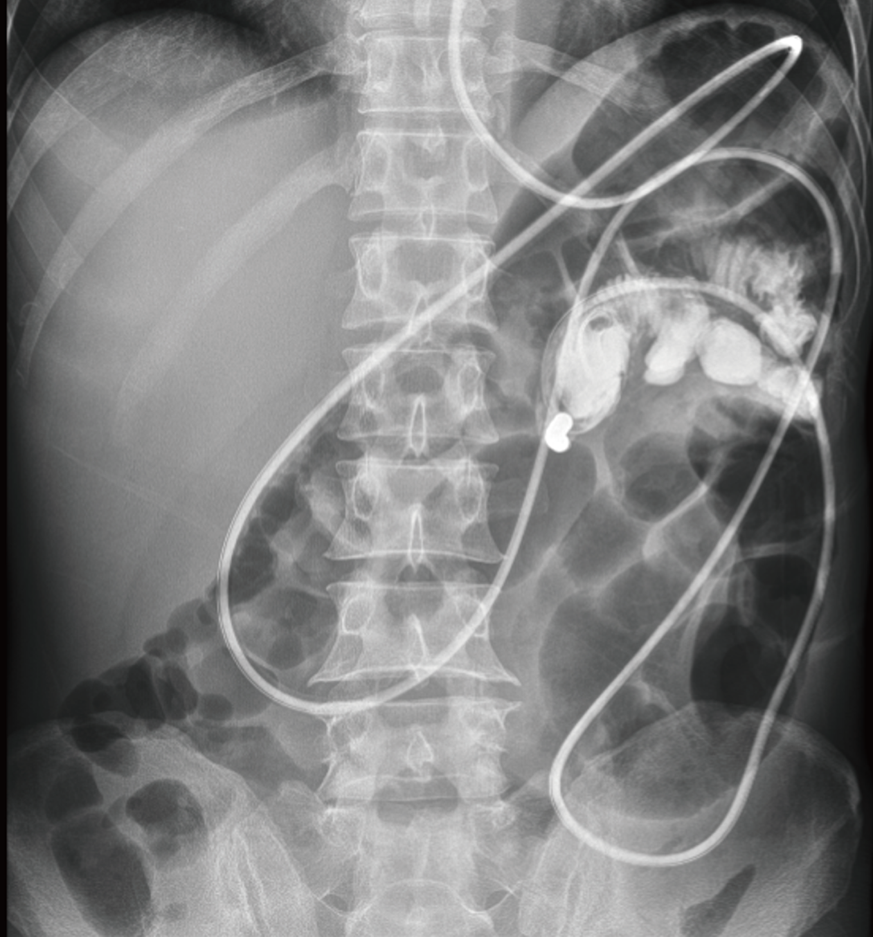
Figure 4 The second fistulogram image.
Gastrointestinal imaging indicated that the catheter had completely crossed the fistula.
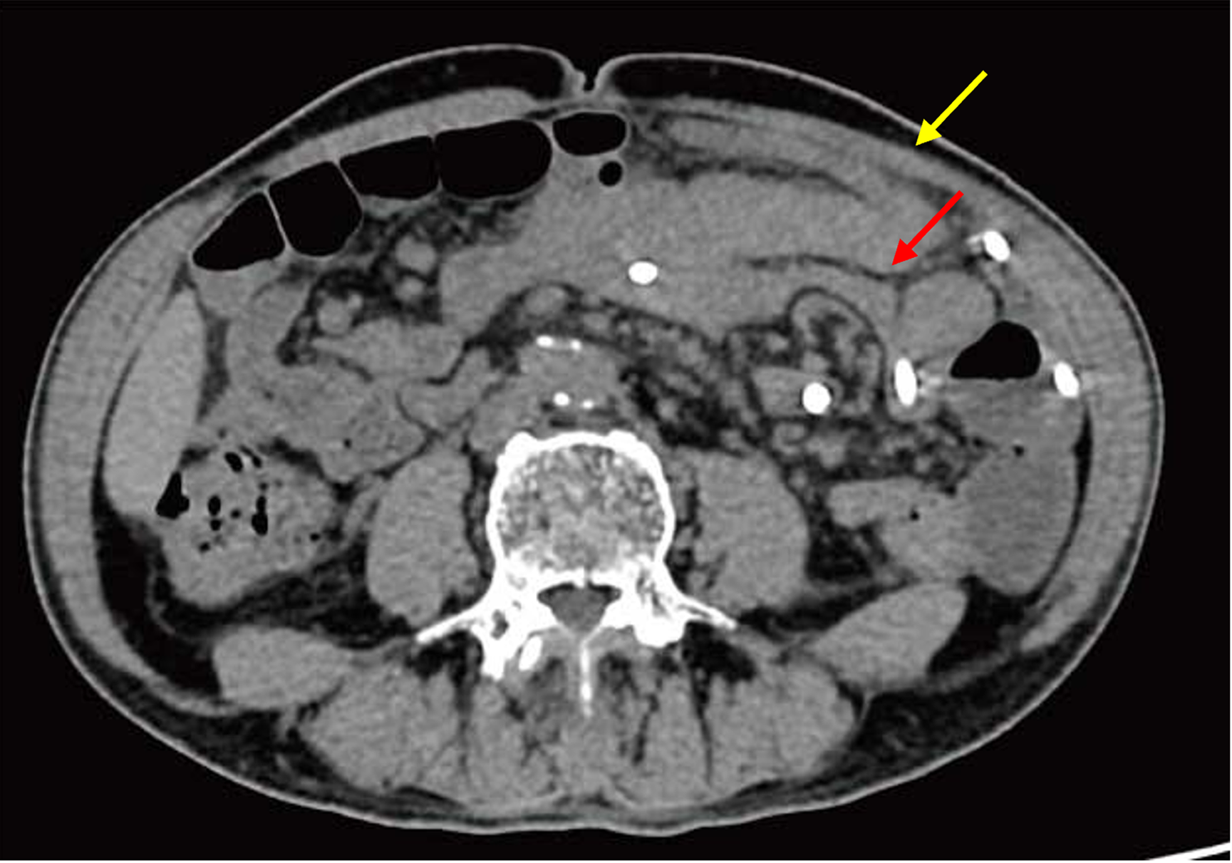
Figure 5 Image of computed tomography scan.
The patient's original fistula has closed at the location pointed out by the red arrow, and the skin fistula has healed as indicated by the yellow arrow.
- Citation: Wang XT, Wang L, Liu AL, Huang JL, Li L, Lu ZX, Mai W. New application of intestinal obstruction catheter in enterocutaneous fistula: A case report. World J Clin Cases 2024; 12(20): 4384-4390
- URL: https://www.wjgnet.com/2307-8960/full/v12/i20/4384.htm
- DOI: https://dx.doi.org/10.12998/wjcc.v12.i20.4384


