©The Author(s) 2024.
World J Clin Cases. May 6, 2024; 12(13): 2275-2280
Published online May 6, 2024. doi: 10.12998/wjcc.v12.i13.2275
Published online May 6, 2024. doi: 10.12998/wjcc.v12.i13.2275
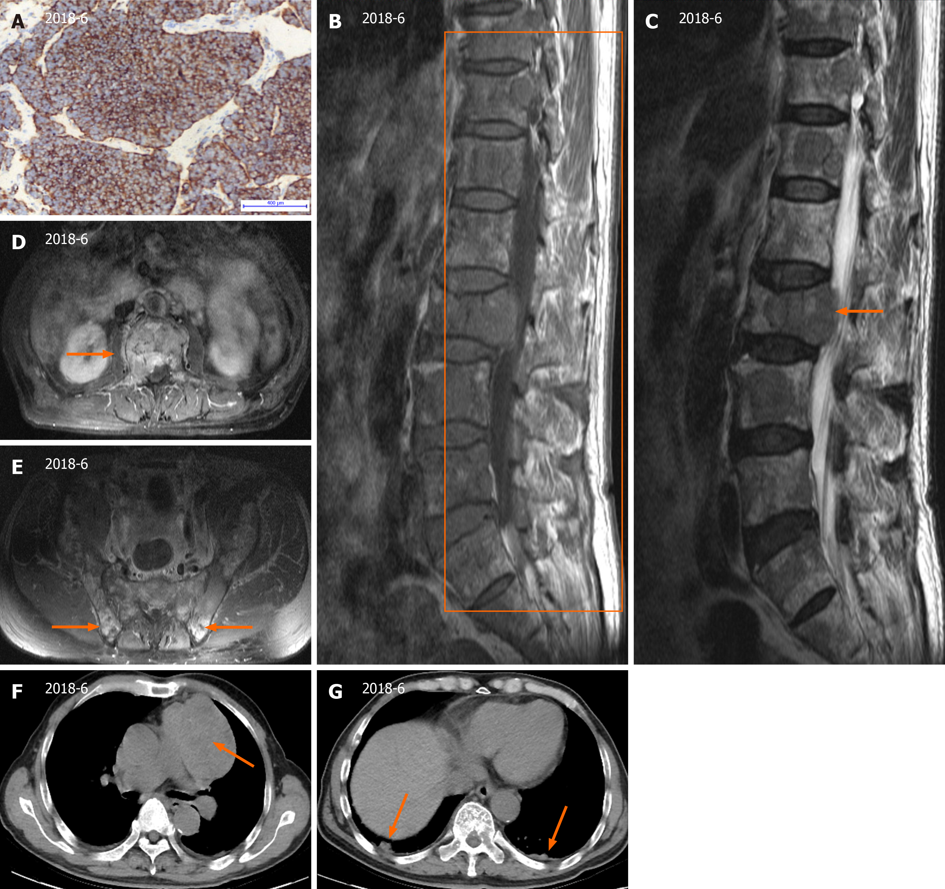
Figure 1 Thymic carcinoid with multiple bone metastases.
A: The original percutaneous needle biopsy of the mediastinal mass showed predominantly single small, round to oval cells with scant cytoplasm and some loose clusters (hematoxylin-eosin staining, × 400); B: Magnetic resonance imaging scans of the lumbar spine showed multiple abnormal signals to the lumbar spine, sacrocaudal vertebrae and iliac crest, indicating bone metastasis; C: L2 vertebral compression fracture and L1/2-L5/S1 with degenerative disc disease; D and E: Lumbar vertebrae axial computed tomography (CT) scan showing bone erosion of the T1 vertebra; F and G: CT scan of the thorax showing a calcified anterior mediastinal mass and a nodular shadow in the subpleural area of both lungs.
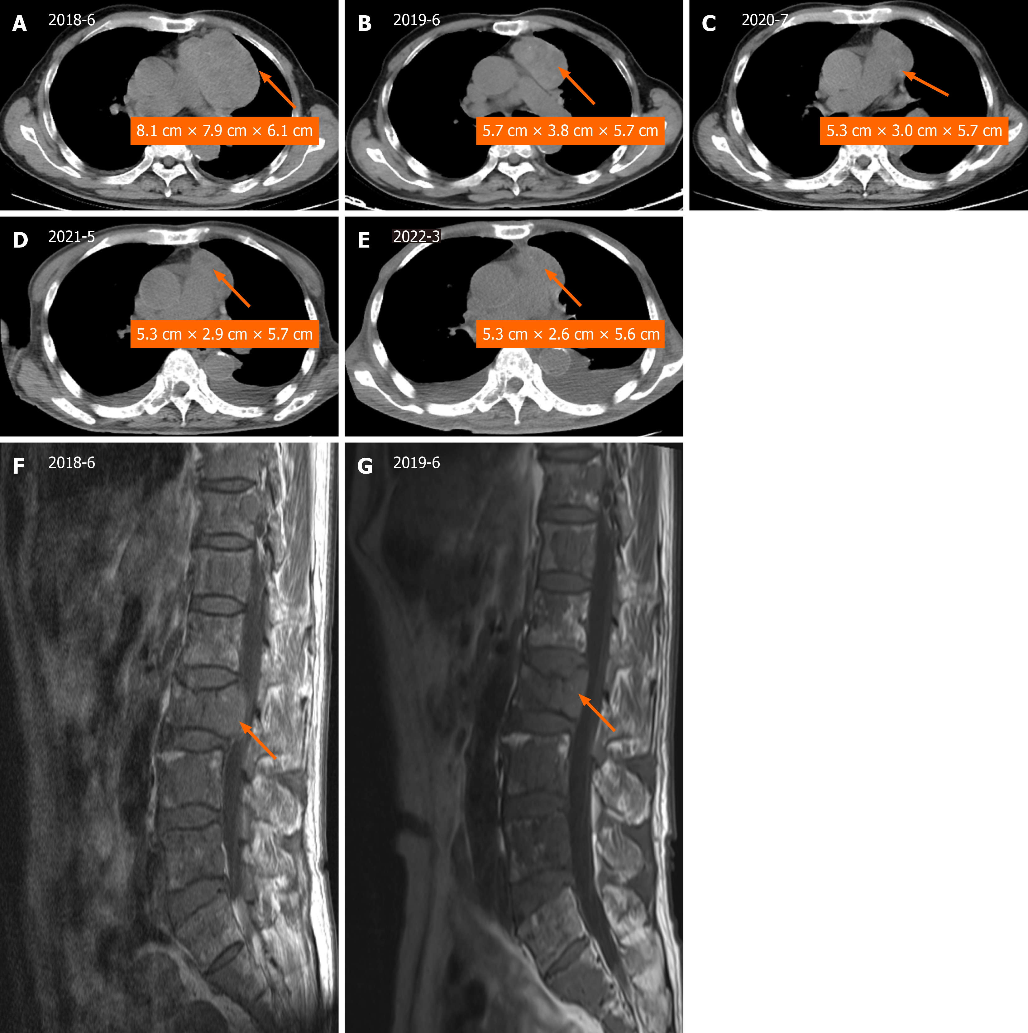
Figure 2 Tumor shrinkage before and after therapy.
A-E: Heterogenous, mediastinal, soft tissue mass observed via computed tomography scan. The mass was measured at the level of branching of the main pulmonary artery; F and G: The vertebral compression fractures improved significantly.
- Citation: Chen CQ, Huang MY, Pan M, Chen QQ, Wei FF, Huang H. Thymic carcinoid with multiple bone metastases: A case report. World J Clin Cases 2024; 12(13): 2275-2280
- URL: https://www.wjgnet.com/2307-8960/full/v12/i13/2275.htm
- DOI: https://dx.doi.org/10.12998/wjcc.v12.i13.2275


