Copyright
©The Author(s) 2023.
World J Clin Cases. Mar 6, 2023; 11(7): 1627-1633
Published online Mar 6, 2023. doi: 10.12998/wjcc.v11.i7.1627
Published online Mar 6, 2023. doi: 10.12998/wjcc.v11.i7.1627
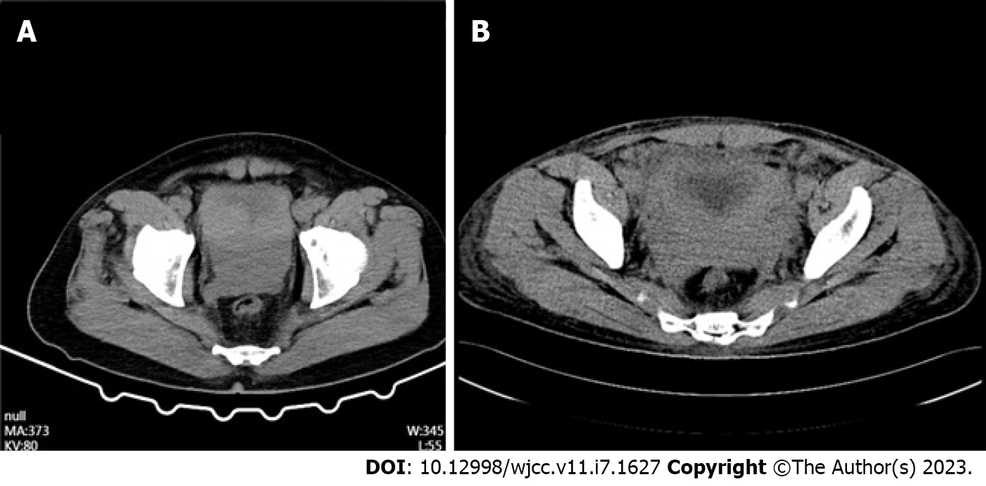
Figure 1 Computed tomography.
A: Significant thickening of the bladder wall with poor demarcation of the prostate-seminiferous gland; B: Thickened bladder wall, poorly defined prostate-seminal gland structure, poorly delineated adjacent pelvic tissue.
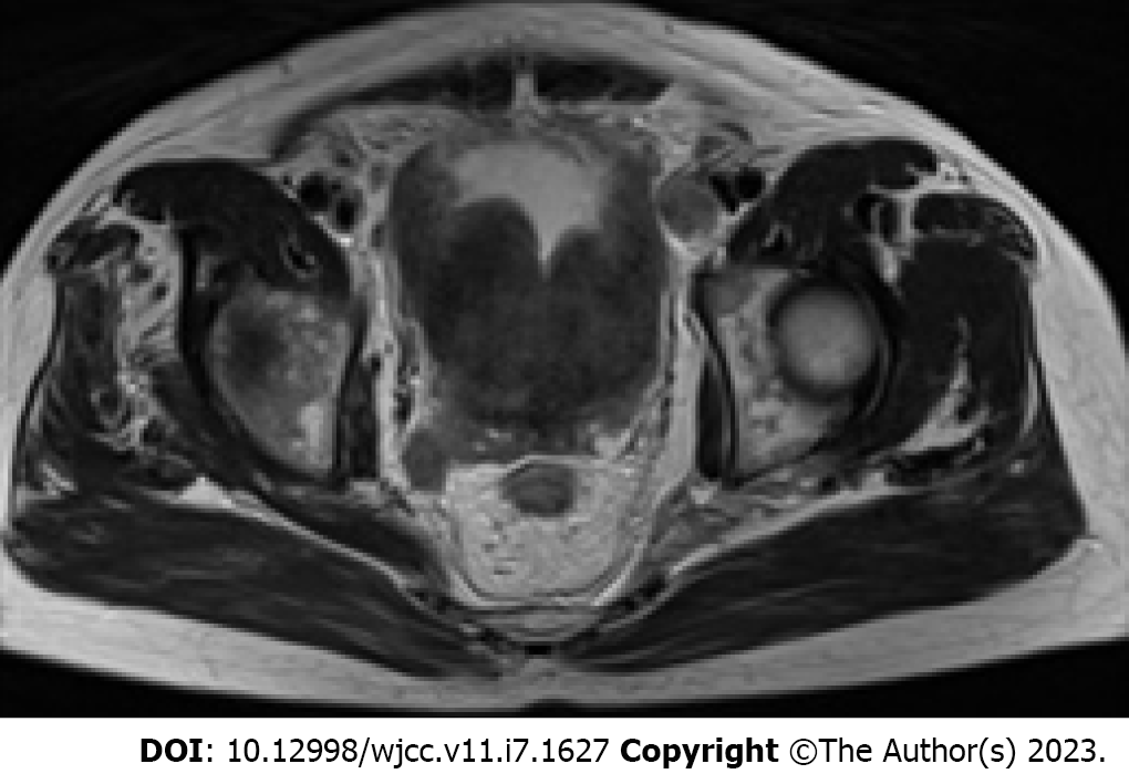
Figure 2 Magnetic resonance imaging.
Abnormal prostate tissue is seen invading the bladder, the bladder wall is thickened, slightly high signal is seen and the surrounding lymph nodes are enlarged.
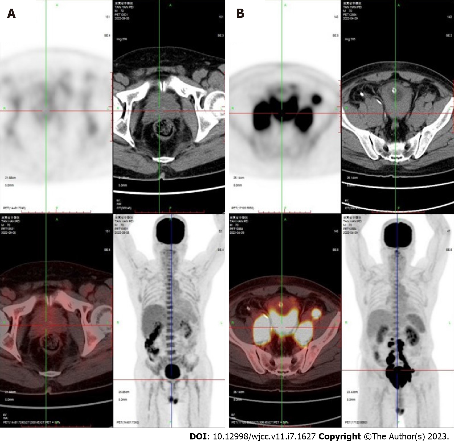
Figure 3 Positron emission tomography/computed tomography imaging before and after treatment.
A: Significantly reduced abnormal prostate hypermetabolic signals after treatment; B: Multiple abnormal signals before treatment.
- Citation: Chen TF, Lin WL, Liu WY, Gu CM. Prostate lymphoma with renal obstruction; reflections on diagnosis and treatment: Two case reports. World J Clin Cases 2023; 11(7): 1627-1633
- URL: https://www.wjgnet.com/2307-8960/full/v11/i7/1627.htm
- DOI: https://dx.doi.org/10.12998/wjcc.v11.i7.1627


