©The Author(s) 2023.
World J Clin Cases. Mar 6, 2023; 11(7): 1607-1614
Published online Mar 6, 2023. doi: 10.12998/wjcc.v11.i7.1607
Published online Mar 6, 2023. doi: 10.12998/wjcc.v11.i7.1607
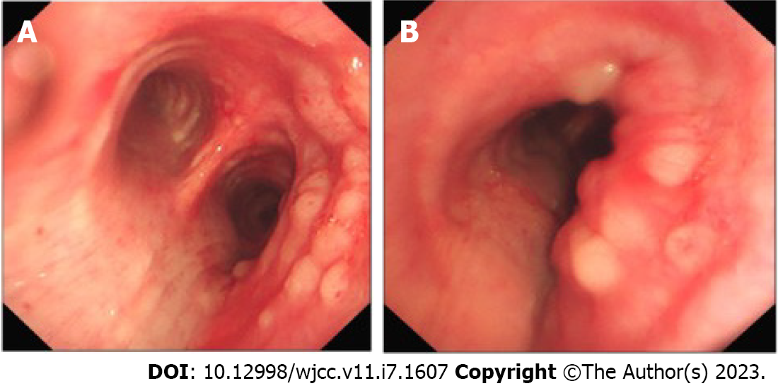
Figure 1 Fiberoptic bronchoscopy.
A: Fiberoptic bronchoscopy image at the tracheal carina; B: Fiberoptic bronchoscopy image at the right main bronchus.
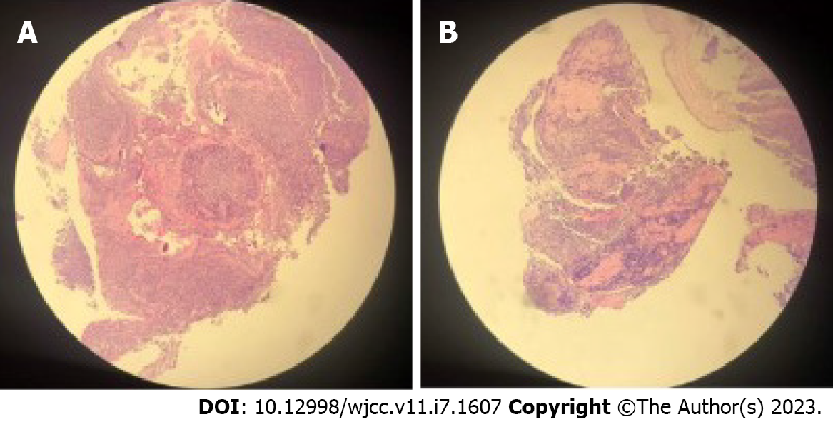
Figure 2 Hematoxylin-eosin staining (H&E) photomicrograph.
A: Part1; B: Part2.
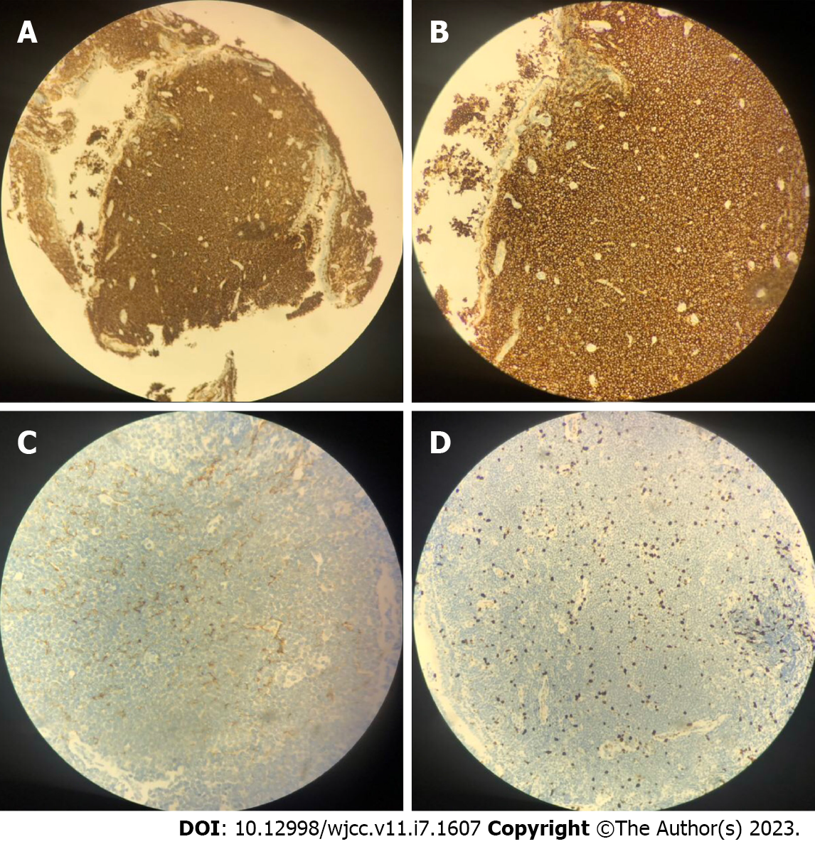
Figure 3 Immunohistochemical stain.
A and B: CD20; C: CD21; D: Ki-67.
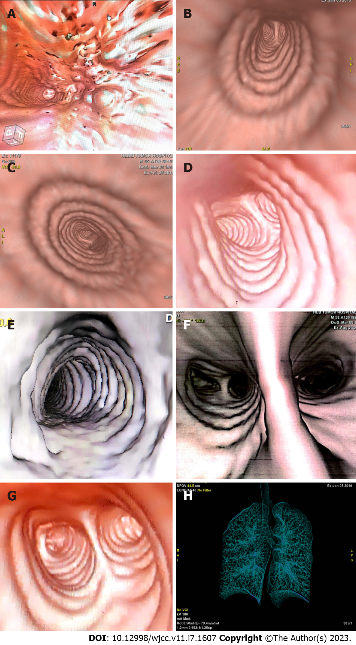
Figure 4 The images of computed tomography virtual bronchoscopy.
A: The image of computed tomography virtual bronchoscopy (CTVB) before radiotherapy (RT); B: CTVB after RT; C: CTVB 1.5 mo after RT; D: CTVB 6 mo after RT; E: CTVB 1.5 years after RT; F: CTVB 2.5 years after RT; G: CTVB 3.5 years after RT; H: Displayed range of CTVB.
- Citation: Zhen CJ, Zhang P, Bai WW, Song YZ, Liang JL, Qiao XY, Zhou ZG. Mucosa-associated lymphoid tissue lymphoma of the trachea treated with radiotherapy: A case report. World J Clin Cases 2023; 11(7): 1607-1614
- URL: https://www.wjgnet.com/2307-8960/full/v11/i7/1607.htm
- DOI: https://dx.doi.org/10.12998/wjcc.v11.i7.1607


