©The Author(s) 2023.
World J Clin Cases. Mar 6, 2023; 11(7): 1586-1592
Published online Mar 6, 2023. doi: 10.12998/wjcc.v11.i7.1586
Published online Mar 6, 2023. doi: 10.12998/wjcc.v11.i7.1586
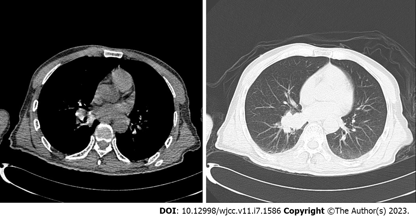
Figure 1 Chest computed tomography.
Soft tissue cluster shadows near the lower left of the right lung.
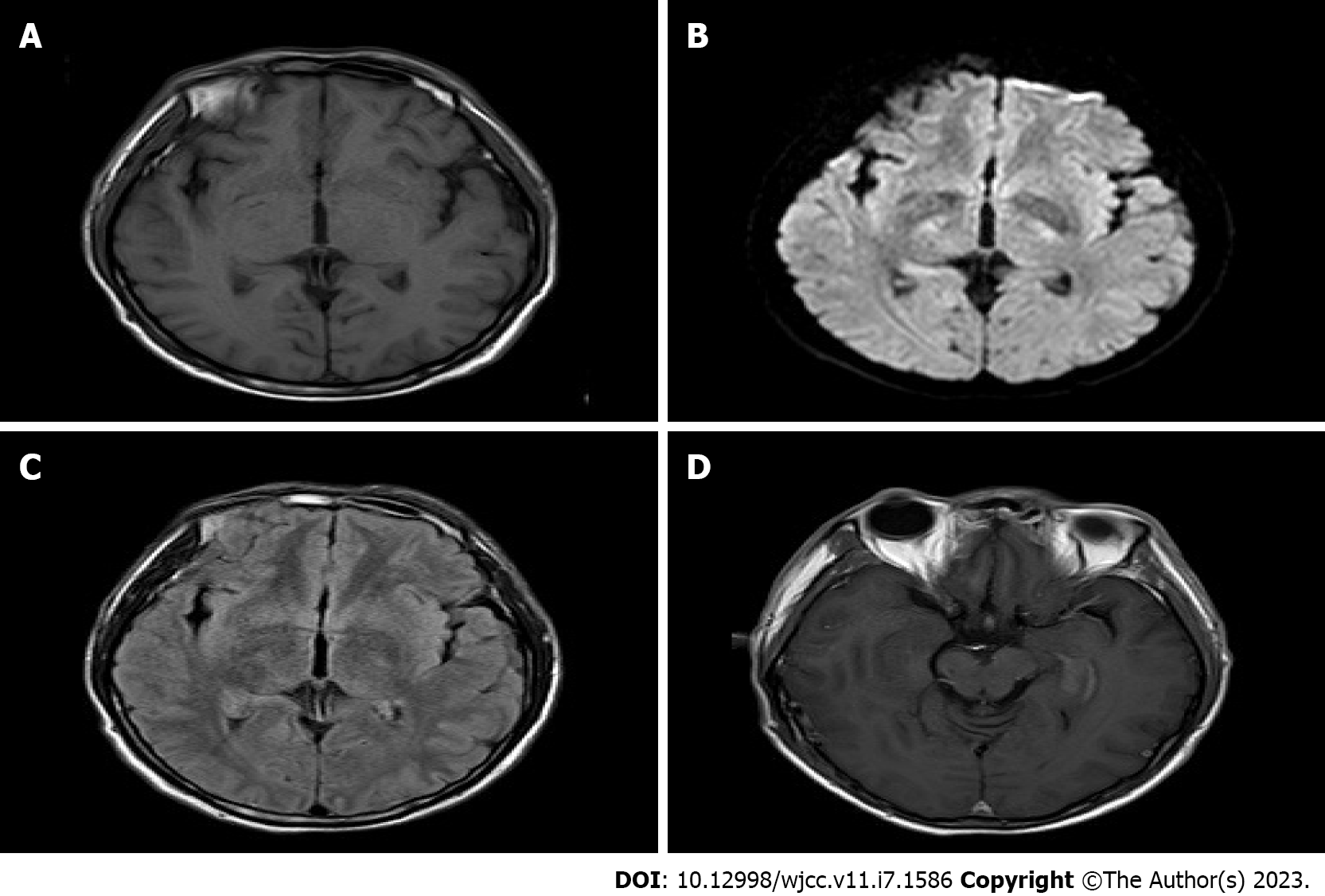
Figure 2 Head magnetic resonance imaging.
A and B: T1 and diffusion-weighted imaging: no abnormal signal; C and D Head magnetic resonance imaging T2 fluid attenuated inversion recovery and Strengthen: the left hippocampus was thickened, and there was a slightly higher signal.
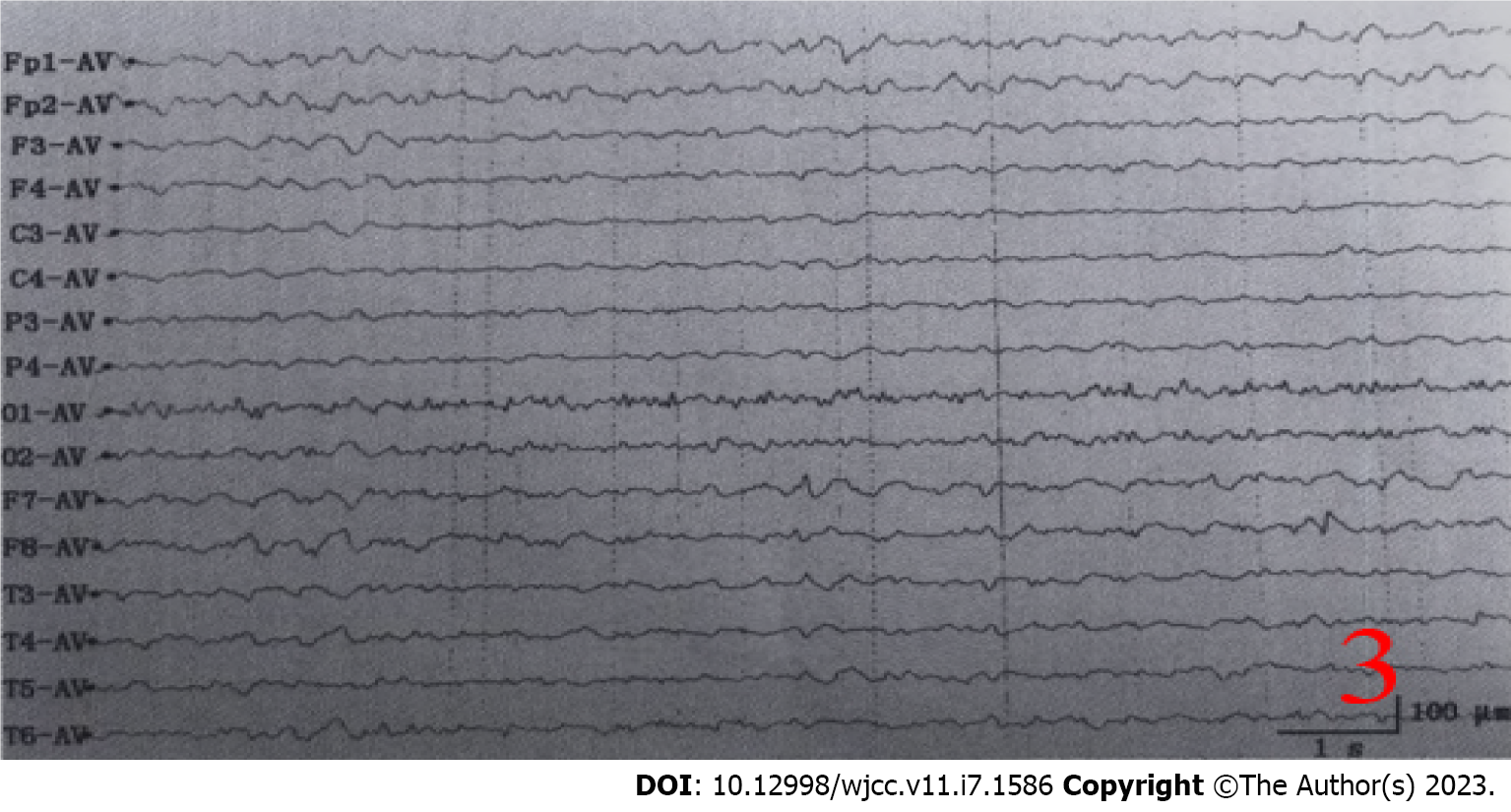
Figure 3 Electroencephalogram.
The patient showed the background was diffuse low-medium amplitude irregular slow wave, with no obvious asymmetry on both sides, and the baseline was unstable.
- Citation: Huang P, Xu M. Four kinds of antibody positive paraneoplastic limbic encephalitis: A rare case report. World J Clin Cases 2023; 11(7): 1586-1592
- URL: https://www.wjgnet.com/2307-8960/full/v11/i7/1586.htm
- DOI: https://dx.doi.org/10.12998/wjcc.v11.i7.1586


