©The Author(s) 2023.
World J Clin Cases. Mar 6, 2023; 11(7): 1576-1585
Published online Mar 6, 2023. doi: 10.12998/wjcc.v11.i7.1576
Published online Mar 6, 2023. doi: 10.12998/wjcc.v11.i7.1576
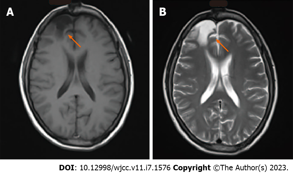
Figure 1 Radiological follow-up 1 year after surgery.
A: T1-weighted and B: T-2 weighted magnetic resonance images. Arrows indicate the site of the pilocytic astrocytoma, which had been previously resected.
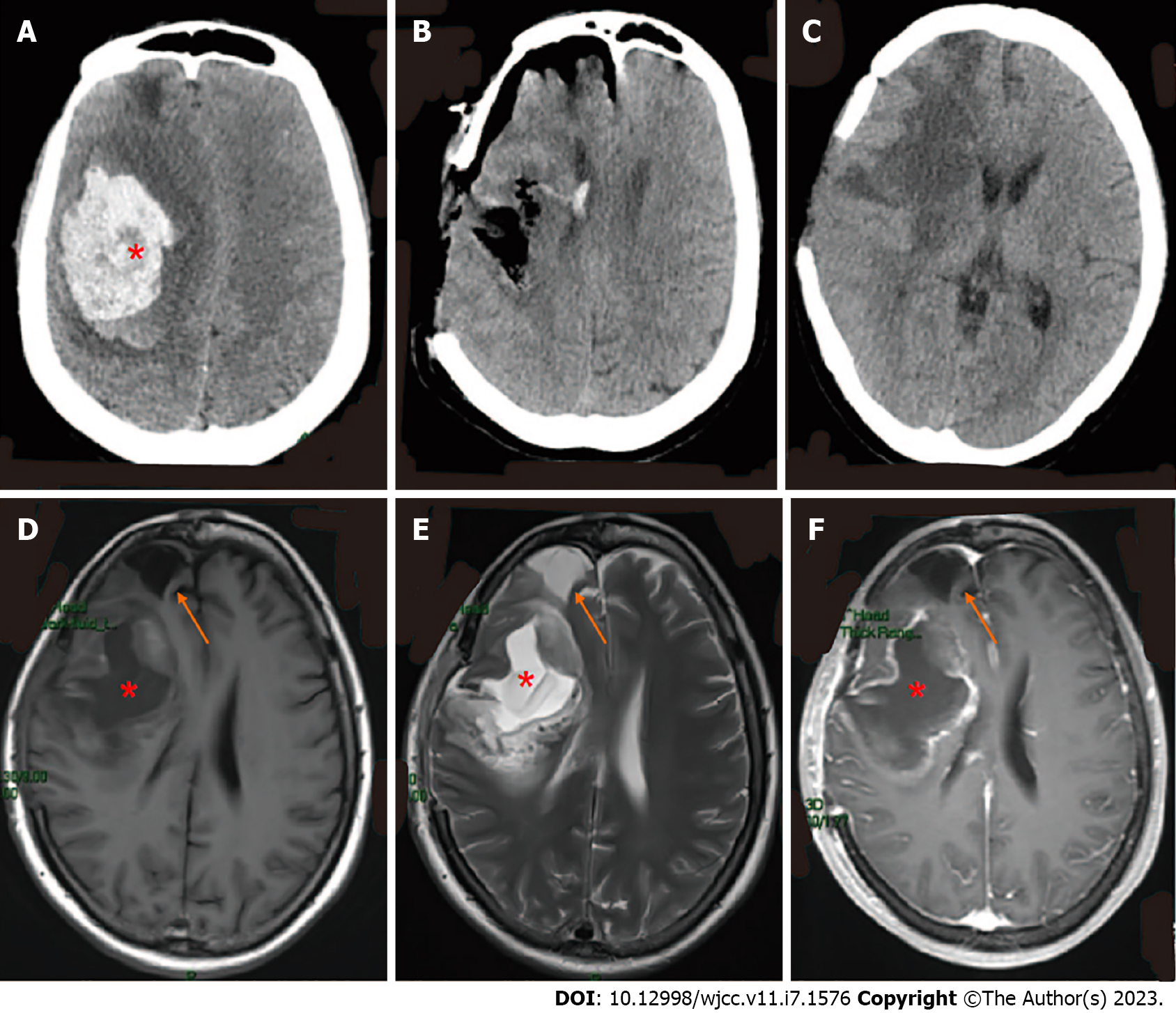
Figure 2 Computed tomography and magnetic resonance imaging throughout admission.
Computed tomography demonstrated A: Right basal ganglia hemorrhage and cerebral herniation at admission; B: Immediate postoperative changes; C: Brain at postoperative day 11; D: T1-weighted and E: T2-weighted cranial magnetic resonance imaging; F: T1-weighted MRI with gadolinium showed right frontal lobe and basal ganglia with marginal enhancement, postoperative day 11. Asterisks (*) indicate hemorrhage; arrows indicate right frontal lobe residual cavity (appears the same as it did 25 years ago).
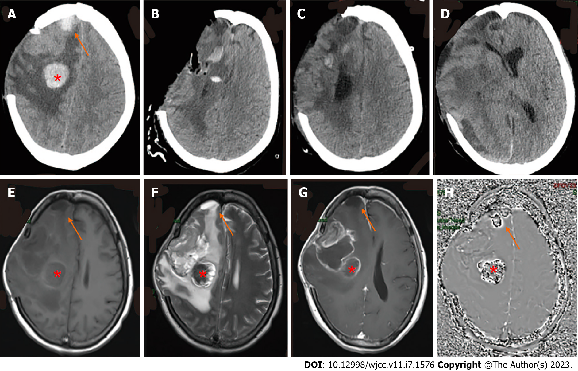
Figure 3 Computed tomography and magnetic resonance imaging with malignant progression of glioma.
A: Computed tomography (CT) showed a new cerebral hemorrhage in the right craniocerebral previous operation site and in the frontal lobe; B: CT after resection of the right frontotemporal parietal lesion plus extended flap decompression; C: On the 23rd d after surgery, the tumor was again observed in the intracranial surgical area and was found to be enlarged; D: On the 50th d after the operation, CT showed rapid tumor growth accompanied by brain herniation; E: T1-weighted, F: T2-weighted, and G: T1-weighted with gadolinium magnetic resonance imaging with the presence of a residual cavity in the right frontoparietal lobe and basal ganglia with marginal enhancement; H: Susceptibility-weighted imaging identifies hemorrhage of frontal lobe and previous operation site. The red asterisk (*) indicates a new basal ganglia hemorrhage; arrows indicate hemorrhage at the right frontal lobe which was identified as pilocytic astrocytoma 25 years previously.
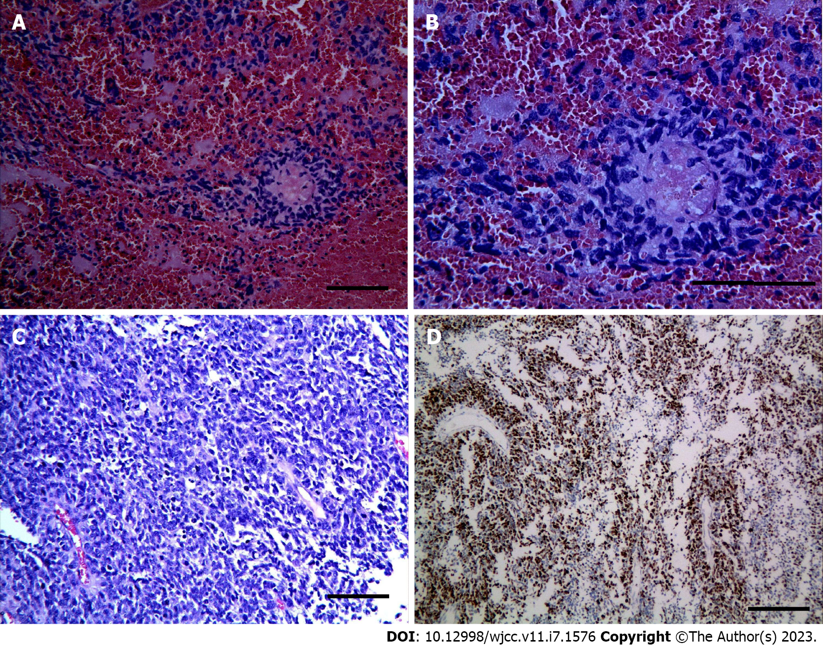
Figure 4 Histology and immunohistochemistry of tumor.
A-C: Photomicrographs of tumor stained with hematoxylin and eosin showed diffusely arranged round cells with perinuclear haloes, akin to those seen in oligodendroglioma (magnification A: 20×; B: 40×; C: 20×); D: The tumor cells demonstrated a proliferation index of 70% by immunohistochemical staining for Ki-67 (magnification: 20×).
- Citation: Xu EX, Lu SY, Chen B, Ma XD, Sun EY. Manifestation of the malignant progression of glioma following initial intracerebral hemorrhage: A case report . World J Clin Cases 2023; 11(7): 1576-1585
- URL: https://www.wjgnet.com/2307-8960/full/v11/i7/1576.htm
- DOI: https://dx.doi.org/10.12998/wjcc.v11.i7.1576


