©The Author(s) 2023.
World J Clin Cases. Mar 6, 2023; 11(7): 1488-1497
Published online Mar 6, 2023. doi: 10.12998/wjcc.v11.i7.1488
Published online Mar 6, 2023. doi: 10.12998/wjcc.v11.i7.1488
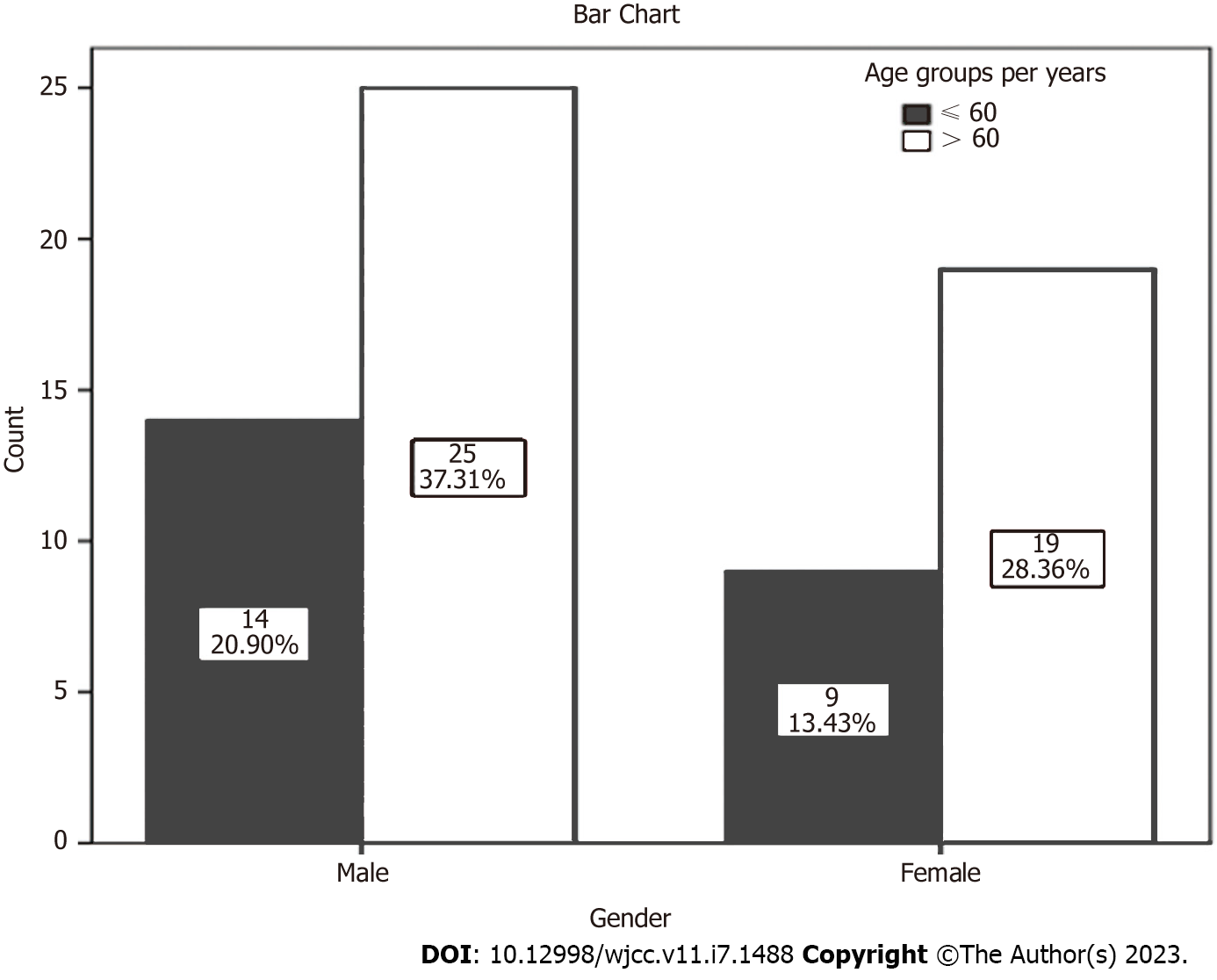
Figure 1 The age and gender distribution of the 67 patients.
P value = 0.750.
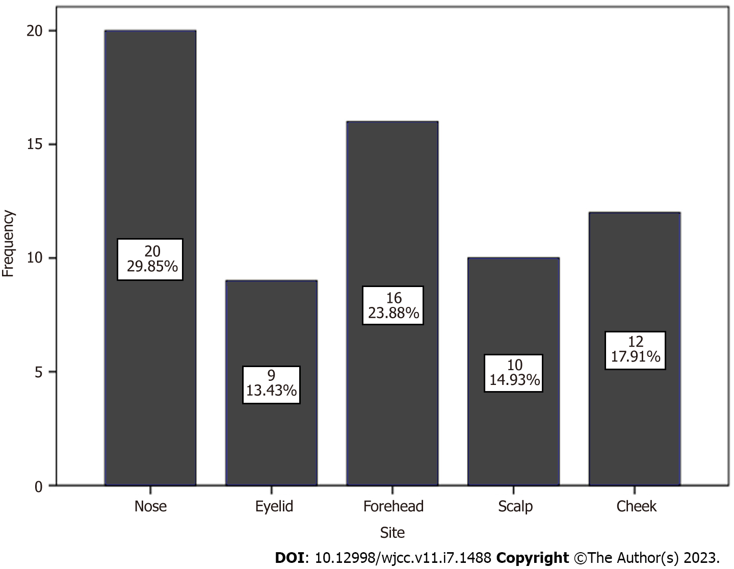
Figure 2 The distribution of the 67 patients according to the site of the facial basal cell carcinoma.
P value = 0.184.
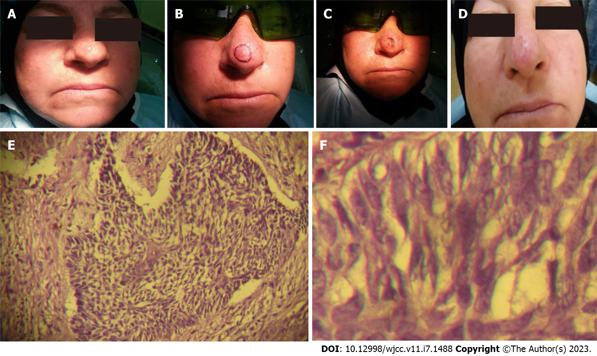
Figure 3 A 60-year-old woman with basal cell carcinoma over the nose.
A: Pigmented lesion; B: Marking the lesion with a safe margin; C: Ablation of the lesion by diode laser; D: Healing by epithelization; E and F: Low (10 ×) and high (40 ×) power of keratotic histopathological type.
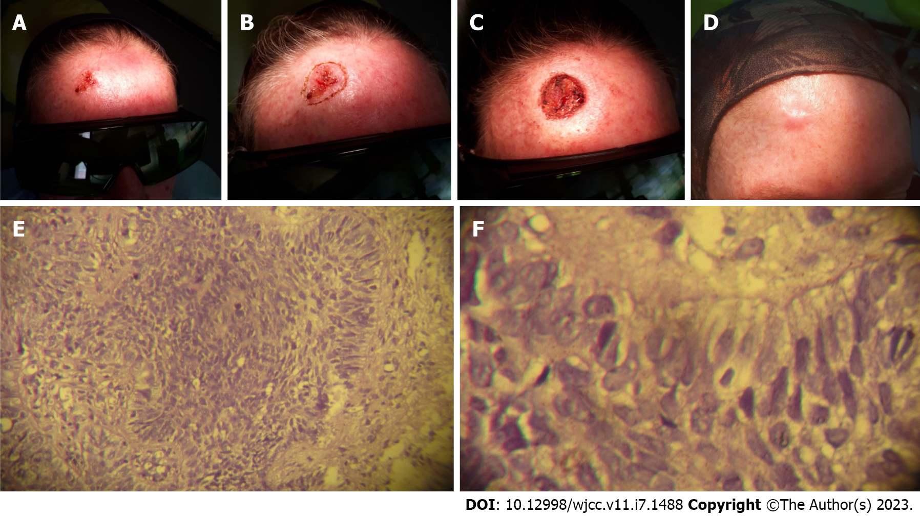
Figure 4 A 45-year-old woman with basal cell carcinoma over the forehead.
A: Ulcerative lesion; B: Barking the lesion with a safe margin; C: Ablation of the lesion by diode laser; D: Healing by epithelization; E and F: Low (10 ×) and high (40 ×) power of solid histopathological type.
- Citation: Khalil AA, Enezei HH, Aldelaimi TN, Al-Ani RM. Facial basal cell carcinoma: A retrospective study of 67 cases. World J Clin Cases 2023; 11(7): 1488-1497
- URL: https://www.wjgnet.com/2307-8960/full/v11/i7/1488.htm
- DOI: https://dx.doi.org/10.12998/wjcc.v11.i7.1488


