©The Author(s) 2023.
World J Clin Cases. Mar 6, 2023; 11(7): 1477-1487
Published online Mar 6, 2023. doi: 10.12998/wjcc.v11.i7.1477
Published online Mar 6, 2023. doi: 10.12998/wjcc.v11.i7.1477
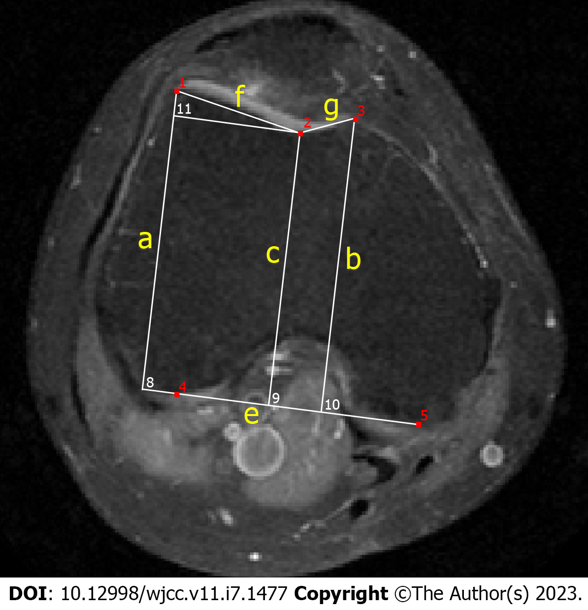
Figure 1 Sample labeling base on axial magnetic resonance imaging.
Trochlear depth was calculated according to the formula [a + b] ÷ 2 - c; asymmetry of the facet length was expressed as [g ÷ f] × 100%; lateral trochlear inclination is the angle between f and e.
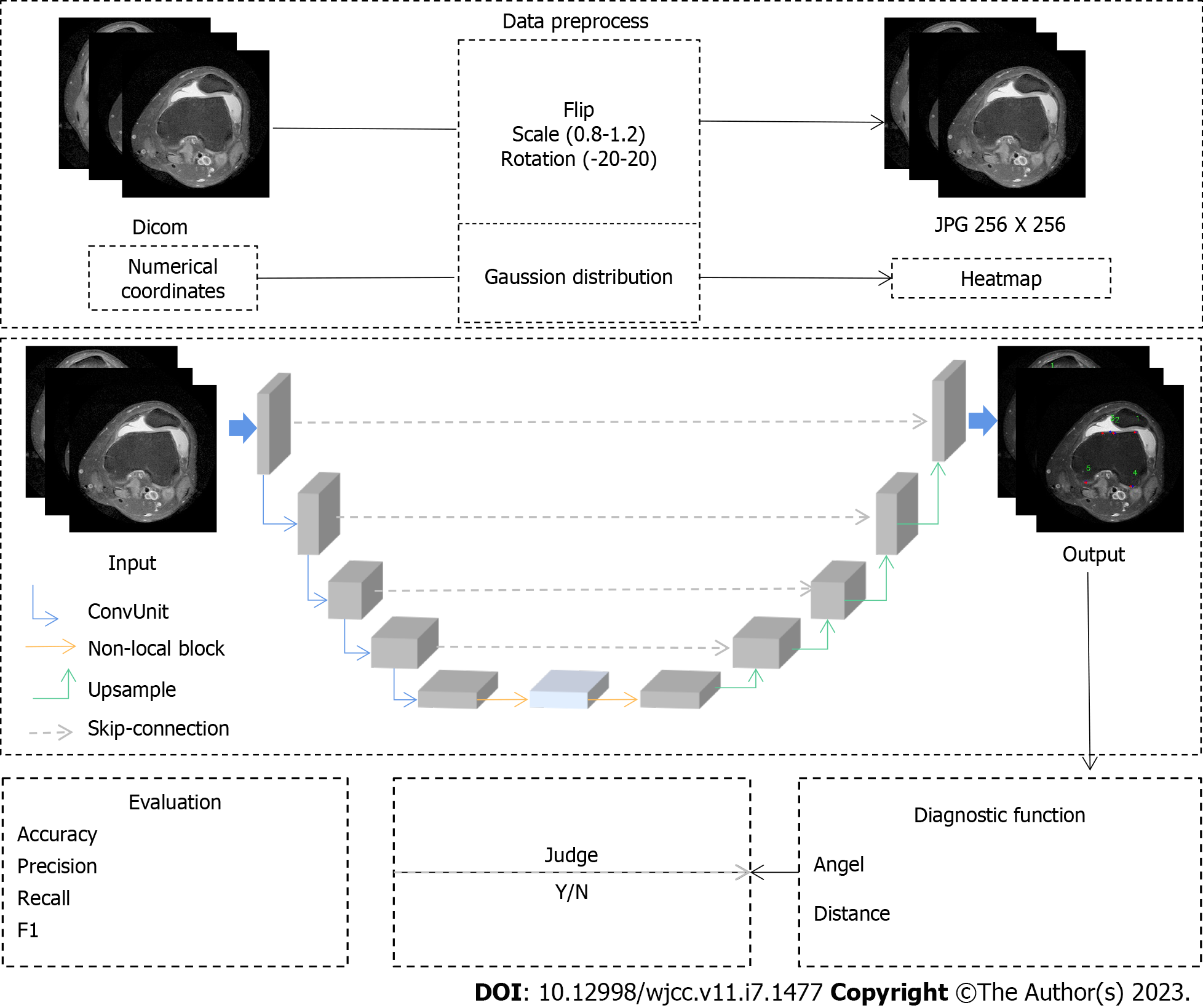
Figure 2 Network structure.
- Citation: Xu SM, Dong D, Li W, Bai T, Zhu MZ, Gu GS. Deep learning-assisted diagnosis of femoral trochlear dysplasia based on magnetic resonance imaging measurements. World J Clin Cases 2023; 11(7): 1477-1487
- URL: https://www.wjgnet.com/2307-8960/full/v11/i7/1477.htm
- DOI: https://dx.doi.org/10.12998/wjcc.v11.i7.1477


