©The Author(s) 2023.
World J Clin Cases. Dec 16, 2023; 11(35): 8357-8363
Published online Dec 16, 2023. doi: 10.12998/wjcc.v11.i35.8357
Published online Dec 16, 2023. doi: 10.12998/wjcc.v11.i35.8357
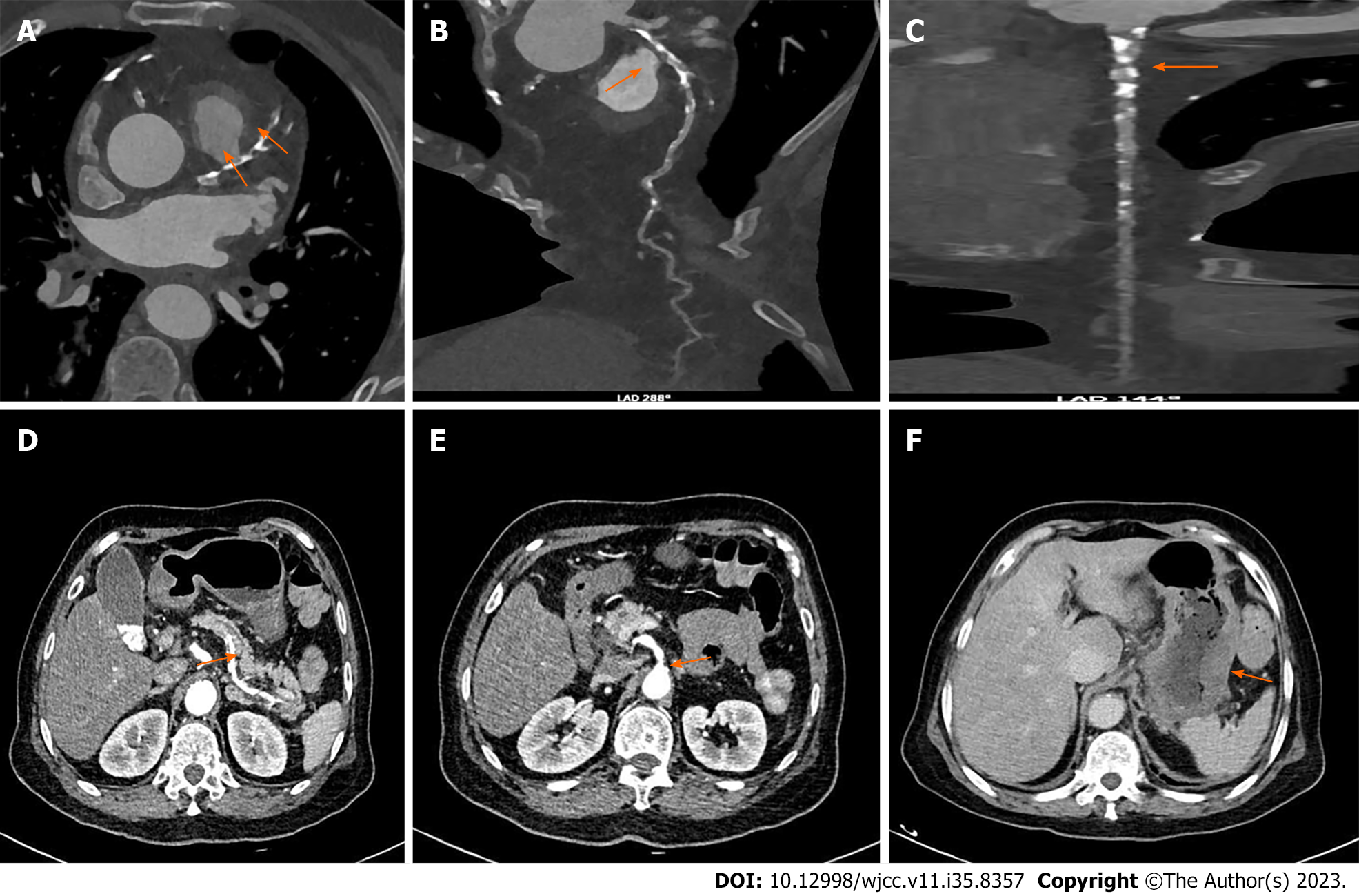
Figure 1 Diffuse mixed plaques in splenic and mesenteric arteries with stenosis of lumen, and atherosclerosis of abdominal aorta and its branches.
A-C: Coronary atherosclerosis, diffuse mixed plaques with moderate to severe stenosis in the proximal and middle segments of the left anterior descending branch (A), multiple calcifications and mixed plaques with mild to moderate stenosis in the proximal and middle segments of the circumflex branch, and multiple localized calcified plaques with mild stenosis in the proximal segments of the right coronary artery (B, C); D and E: Diffuse mixed plaques in the splenic and mesenteric arteries, with stenosis of the lumen, and atherosclerosis of the abdominal aorta and its branches; F: Edema and thickening of the gastric wall.
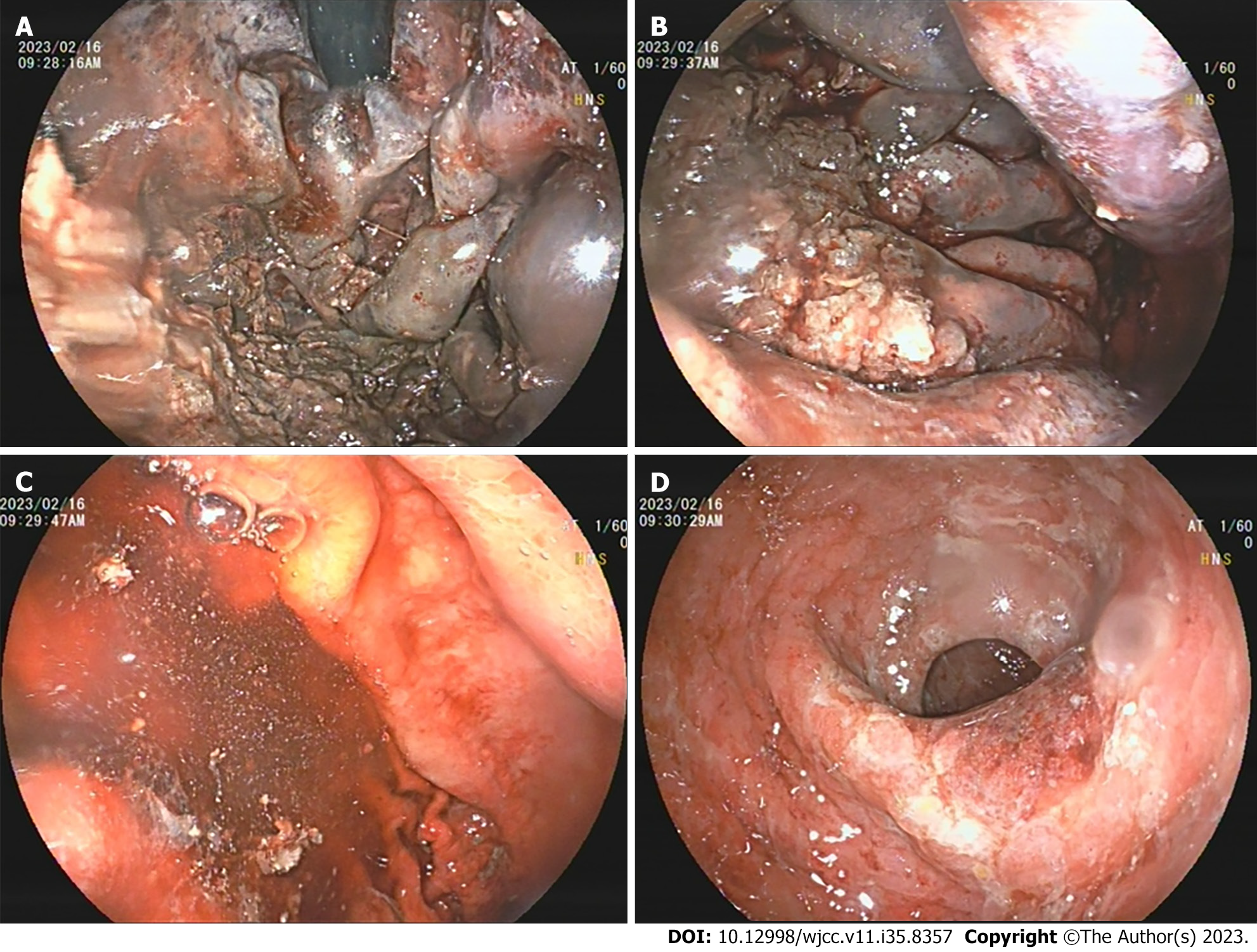
Figure 2 Upper gastrointestinal endoscopy performed on day 2 in hospital.
It showed longitudinal ulcers, multiple irregular ulcers, mucosal edema with redness, erosion, and hemorrhage in the stomach. A: Fundus of stomach; B: Gastric body; C: Gastric body; D: Gastric antrum.
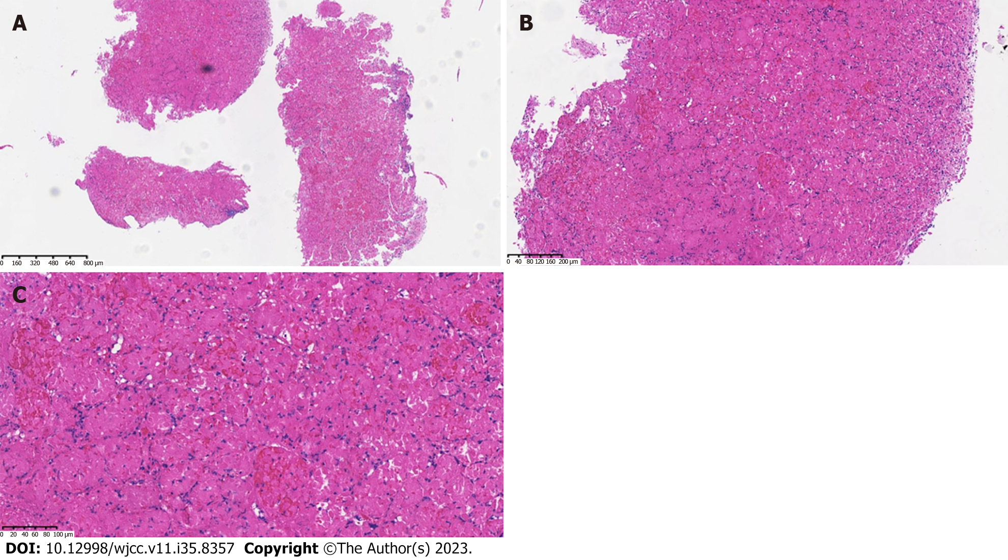
Figure 3 Histopathological findings.
Pathological evaluation of the specimen showed hemorrhage and necrosis of gastric mucosa, and visible residual contour of gastric glands. A: 10 ×; B: 40 ×; C: 100 ×.
- Citation: Wei RY, Zhu JH, Li X, Wu JY, Liu JW. Diffuse arterial atherosclerosis presenting with acute ischemic gastritis: A case report. World J Clin Cases 2023; 11(35): 8357-8363
- URL: https://www.wjgnet.com/2307-8960/full/v11/i35/8357.htm
- DOI: https://dx.doi.org/10.12998/wjcc.v11.i35.8357


