©The Author(s) 2023.
World J Clin Cases. Nov 6, 2023; 11(31): 7570-7582
Published online Nov 6, 2023. doi: 10.12998/wjcc.v11.i31.7570
Published online Nov 6, 2023. doi: 10.12998/wjcc.v11.i31.7570
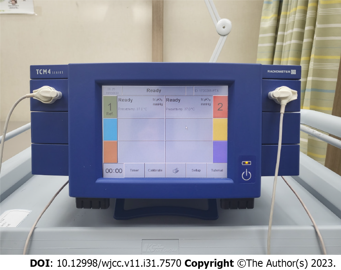
Figure 1 Transcutaneous oxygen pressure measuring instrument (TCM400®).
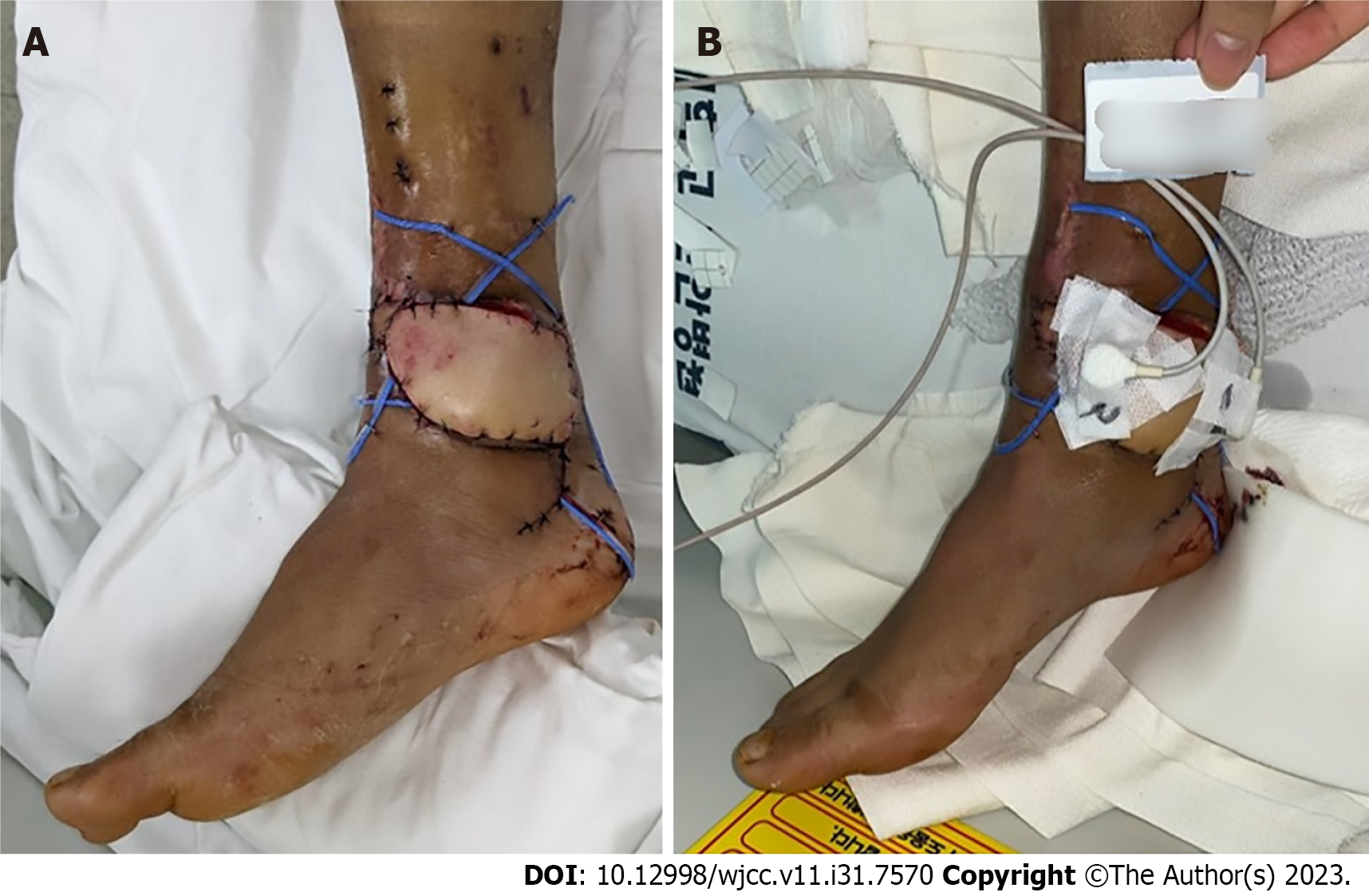
Figure 2 A photographic image of transcutaneous oxygen pressure applied to an anterolateral thigh free flap patient.
A: On postoperative day 1, free flap operation site surviving well; B: Place two probes on the skin perforator site (site marked 1 in the image) and the most distal part of flap (site marked 2 in the image), respectively.
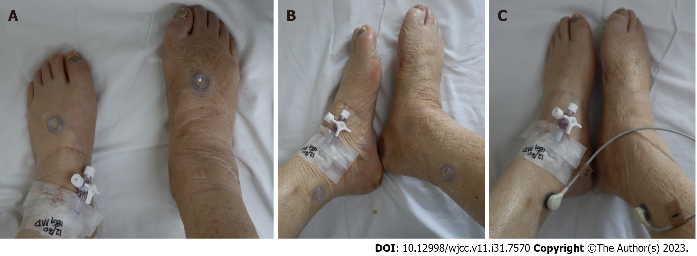
Figure 3 A photographic image of transcutaneous oxygen pressure applied to a diabetic foot ulcer patient without free flap recon
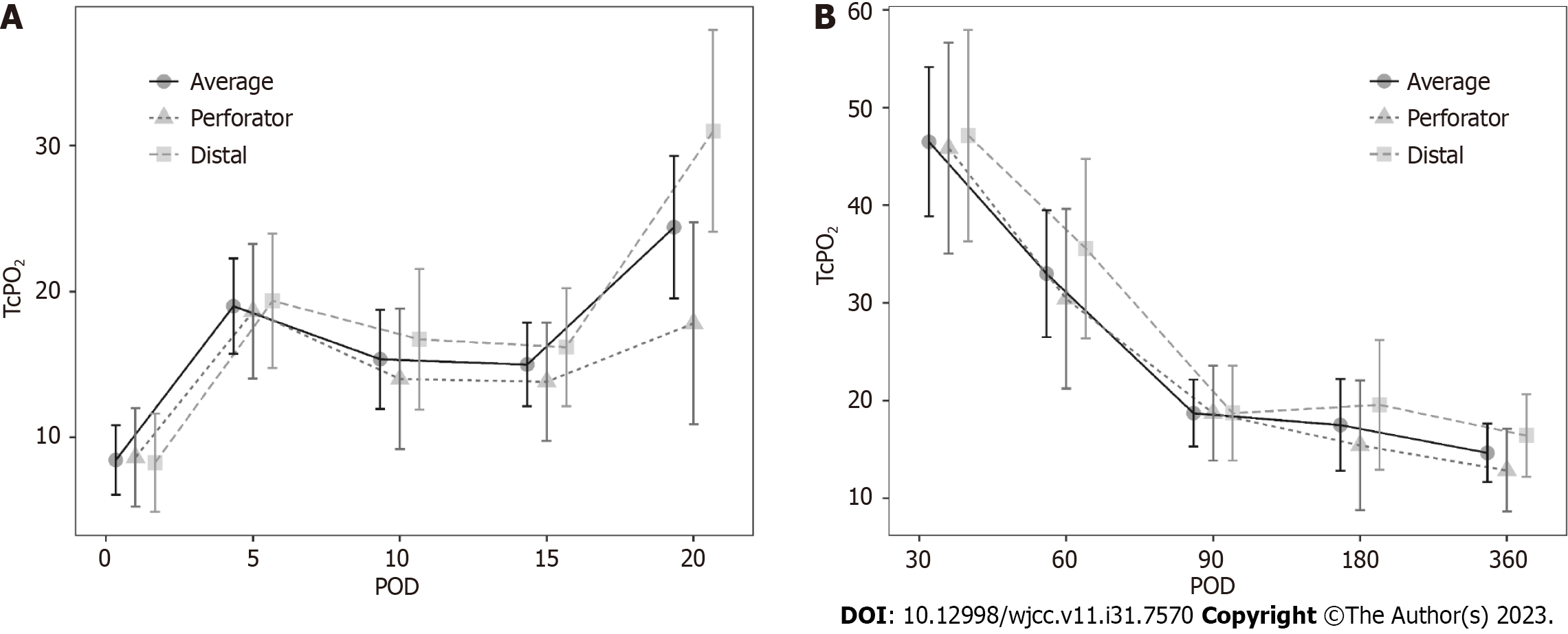
Figure 4 Change in transcutaneous oxygen pressure during the short-term and long-term period.
A: Short-term period. Although the value is low, a gradual increasing pattern is observed; B: Long-term period. Although the value is low, a gradual decreasing trend is observed. POD: Postoperative day.
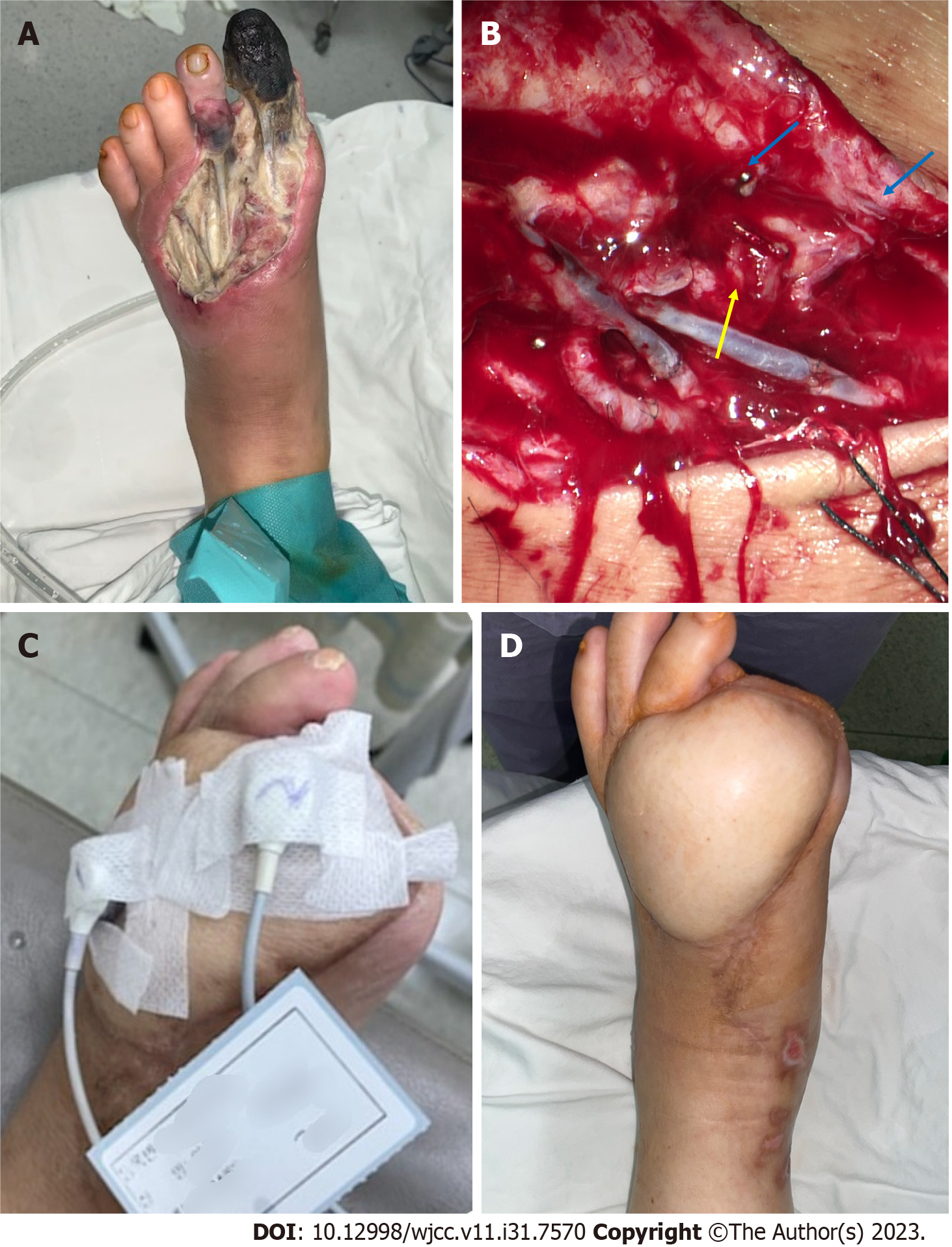
Figure 5 Photographic findings of patient 1.
A: Diabetic foot ulcer (DFU) on the left foot dorsum with necrotic tissue observed before debridement; B: One artery and two vena comitans successfully undergo end-to-end anastomosis (yellow arrow: Artery, blue arrow: Vein); C: Transcutaneous oxygen pressure measured on postoperative day (POD) 30; D: Successfully reconstructed DFU wound on POD 360.
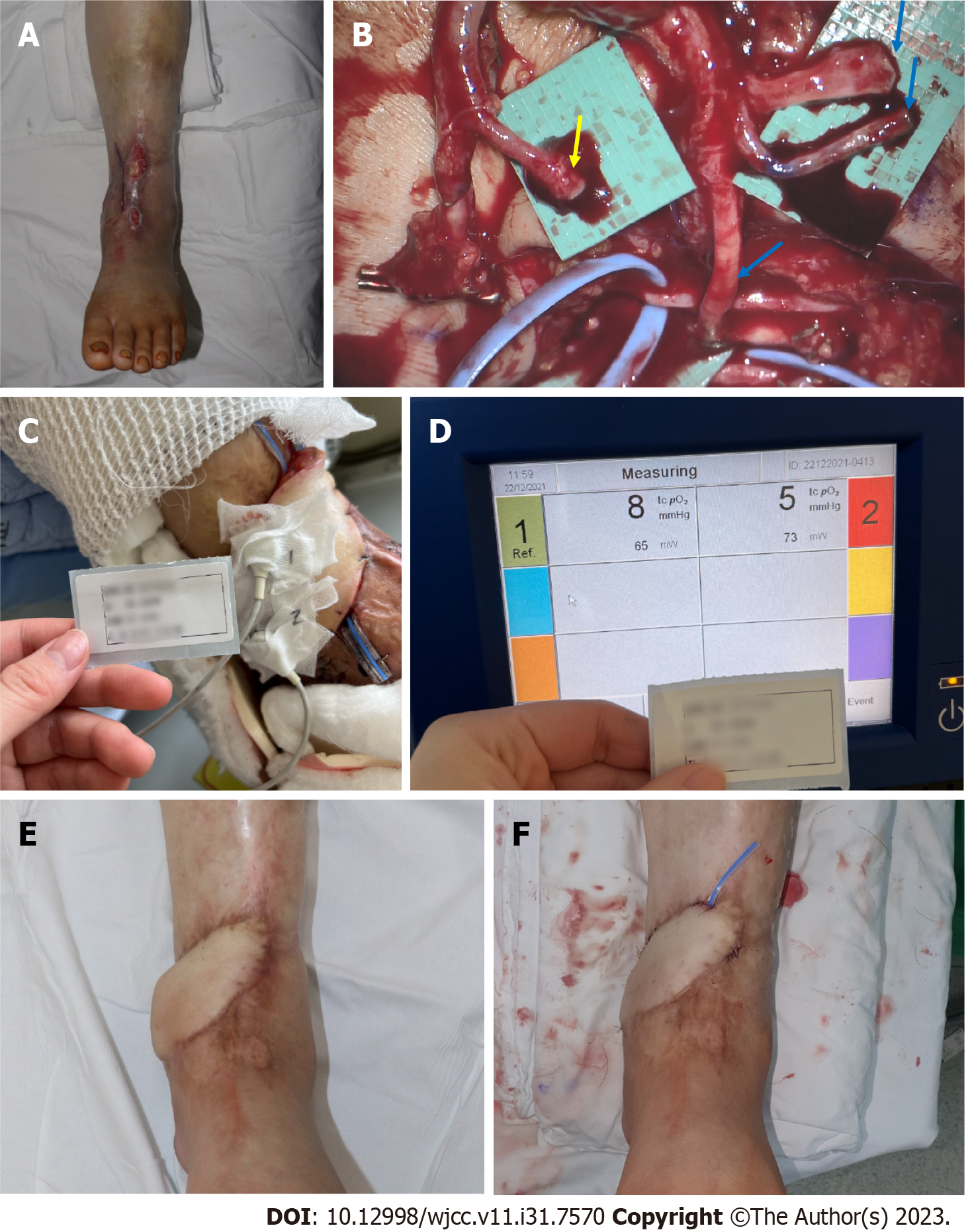
Figure 6 Photographic findings of patient 2.
A: Preoperative findings, tendon, and periosteum exposed; B: The adventitia is stripped from the artery (yellow arrow), and the three veins are anastomosed (blue arrow); C and D: Transcutaneous oxygen pressure measured at postoperative day (POD) 1. Very low values of 8 and 5 mmHg are shown; E: Before flap debulking on POD 180; F: After flap debulking was performed on POD 192.
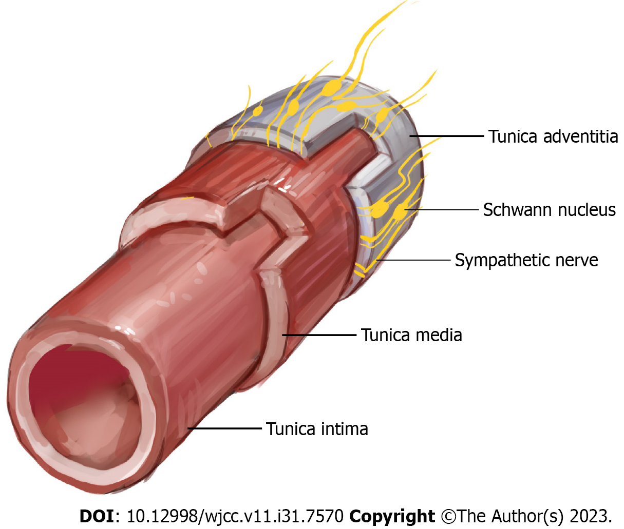
Figure 7 An illustration depicting intima, media, adventitia, and sympathetic innervation in a cross section of a vessel.
Sympathetic nerves are mainly located in the outermost layer, the adventitia.
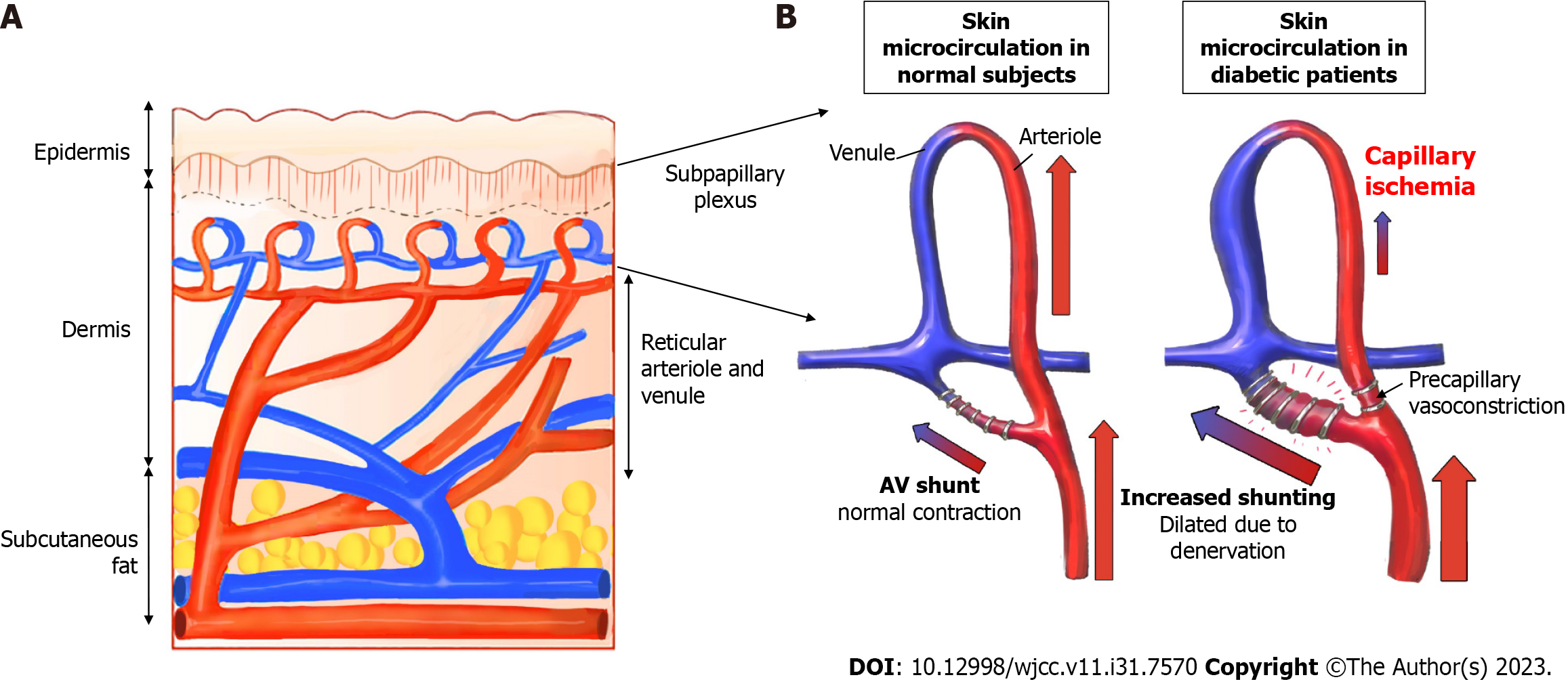
Figure 8 Schematic illustration of the increase in a subpapillary arteriovenous shunt in diabetic patients.
A: Cross-sectional structure of the skin; the subpapillary plexus is located in the lower layer of the epidermis; B: In a patent with diabetes, arteriovenous shunt is increased, and periphery blood flow is reduced.
- Citation: Lee DW, Hwang YS, Byeon JY, Kim JH, Choi HJ. Does the advantage of transcutaneous oximetry measurements in diabetic foot ulcer apply equally to free flap reconstruction? World J Clin Cases 2023; 11(31): 7570-7582
- URL: https://www.wjgnet.com/2307-8960/full/v11/i31/7570.htm
- DOI: https://dx.doi.org/10.12998/wjcc.v11.i31.7570


