Copyright
©The Author(s) 2023.
World J Clin Cases. Jan 26, 2023; 11(3): 709-718
Published online Jan 26, 2023. doi: 10.12998/wjcc.v11.i3.709
Published online Jan 26, 2023. doi: 10.12998/wjcc.v11.i3.709
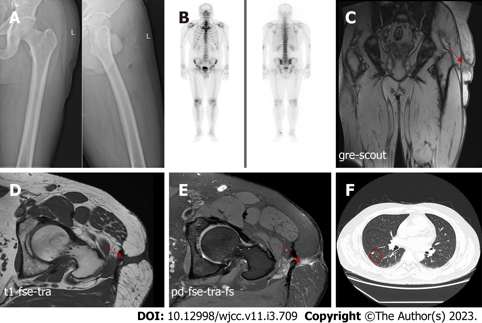
Figure 1 Imaging pictures of the patient.
A: Anteroposterior and lateral radiographs of the left femur are normal; B: Bone single-photon emission computed tomography did not reveal any abnormalities in bone metabolism; C-E: Magnetic resonance imaging of the left hip showing abnormal signals in the soft tissue of the left upper femur and suggesting a chronic abscess with sinus tract formation; F: Computed tomography of the chest showing scattered nodules in both lungs, the largest nodule in the right lung with a length of 8 mm.
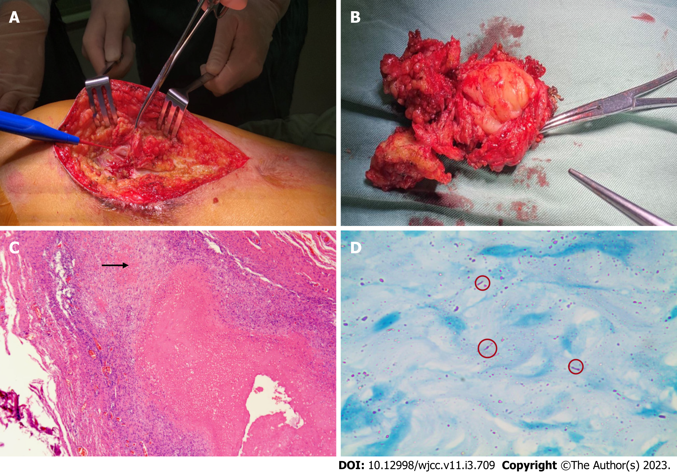
Figure 2 Intraoperative photographs of the left thigh and histopathological examination of resected specimens.
A: Intraoperative photograph; B: Excised specimen (8 cm × 6 cm); C: Granulomas are embedded among the muscle fibers with lymphocyte infiltration and multinucleated giant cell aggregation (× 40); D: The acid-fast bacilli staining test showed scattered, suspicious antacid staining-positive rods (× 1000).
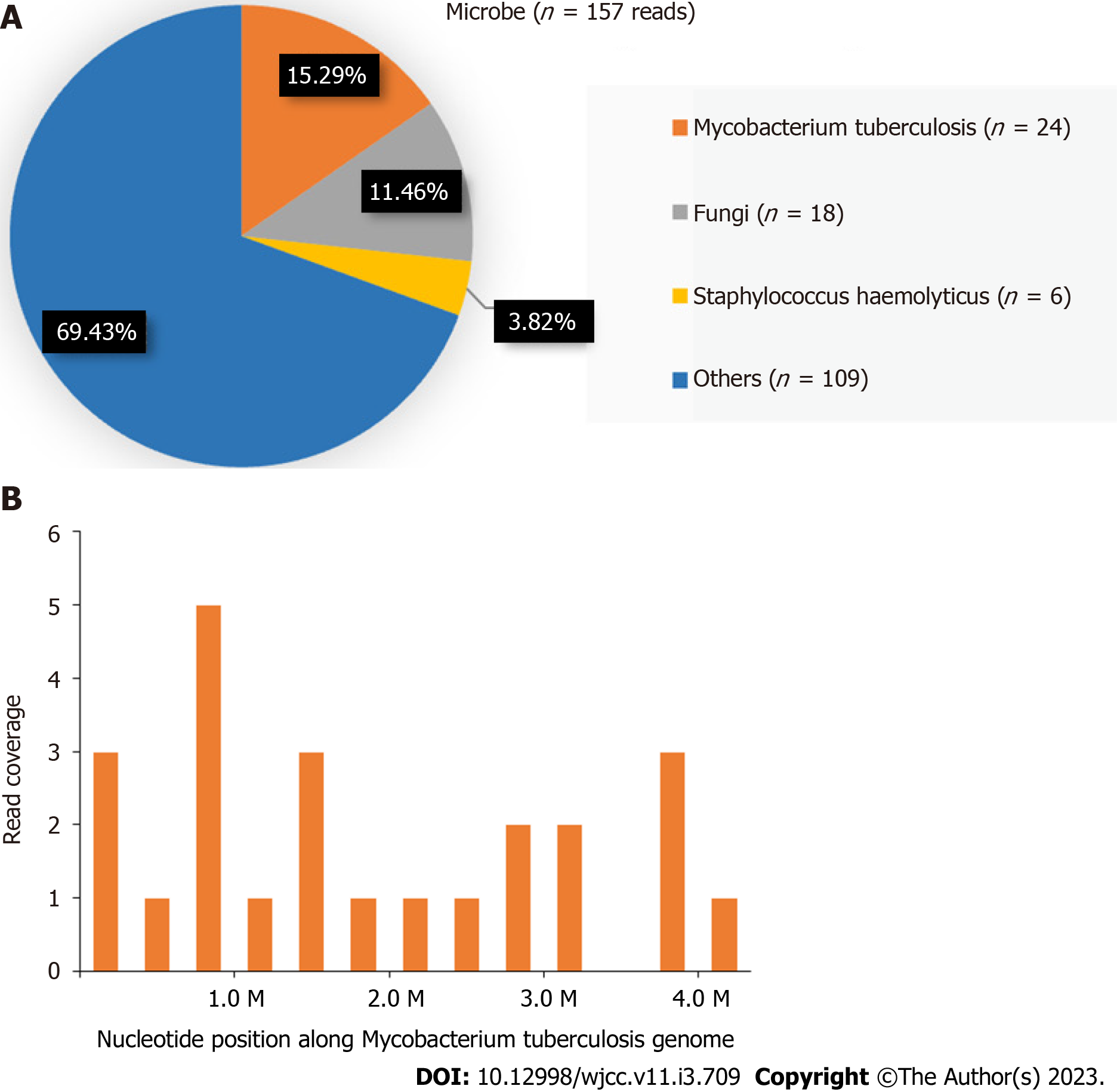
Figure 3 Next-generation sequencing results of surgically resected specimen.
A: Distribution of the sequences detected by next-generation sequencing of surgically excised specimens; B: Twenty-four sequence reads of mycobacterium tuberculosis are observed, with a coverage rate of 0.005%.
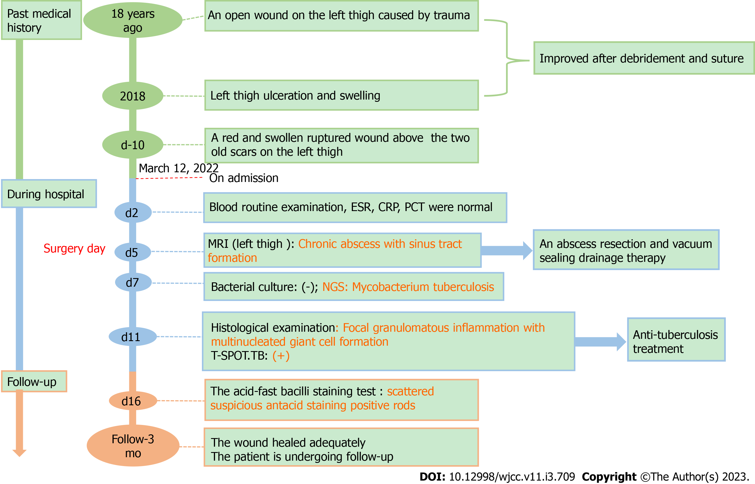
Figure 4 A timeline showing the progress of the disease and the patient’s treatment and follow-up.
ESR: Erythrocyte sedimentation rate; CRP: C-reactive protein; PCT: Procalcitonin; MRI: Magnetic resonance imaging; NGS: Next-generation sequencing.
- Citation: He YG, Huang YH, Yi XL, Qian KL, Wang Y, Cheng H, Hu J, Liu Y. Soft tissue tuberculosis detected by next-generation sequencing: A case report and review of literature. World J Clin Cases 2023; 11(3): 709-718
- URL: https://www.wjgnet.com/2307-8960/full/v11/i3/709.htm
- DOI: https://dx.doi.org/10.12998/wjcc.v11.i3.709


