Copyright
©The Author(s) 2023.
World J Clin Cases. Oct 16, 2023; 11(29): 7248-7252
Published online Oct 16, 2023. doi: 10.12998/wjcc.v11.i29.7248
Published online Oct 16, 2023. doi: 10.12998/wjcc.v11.i29.7248
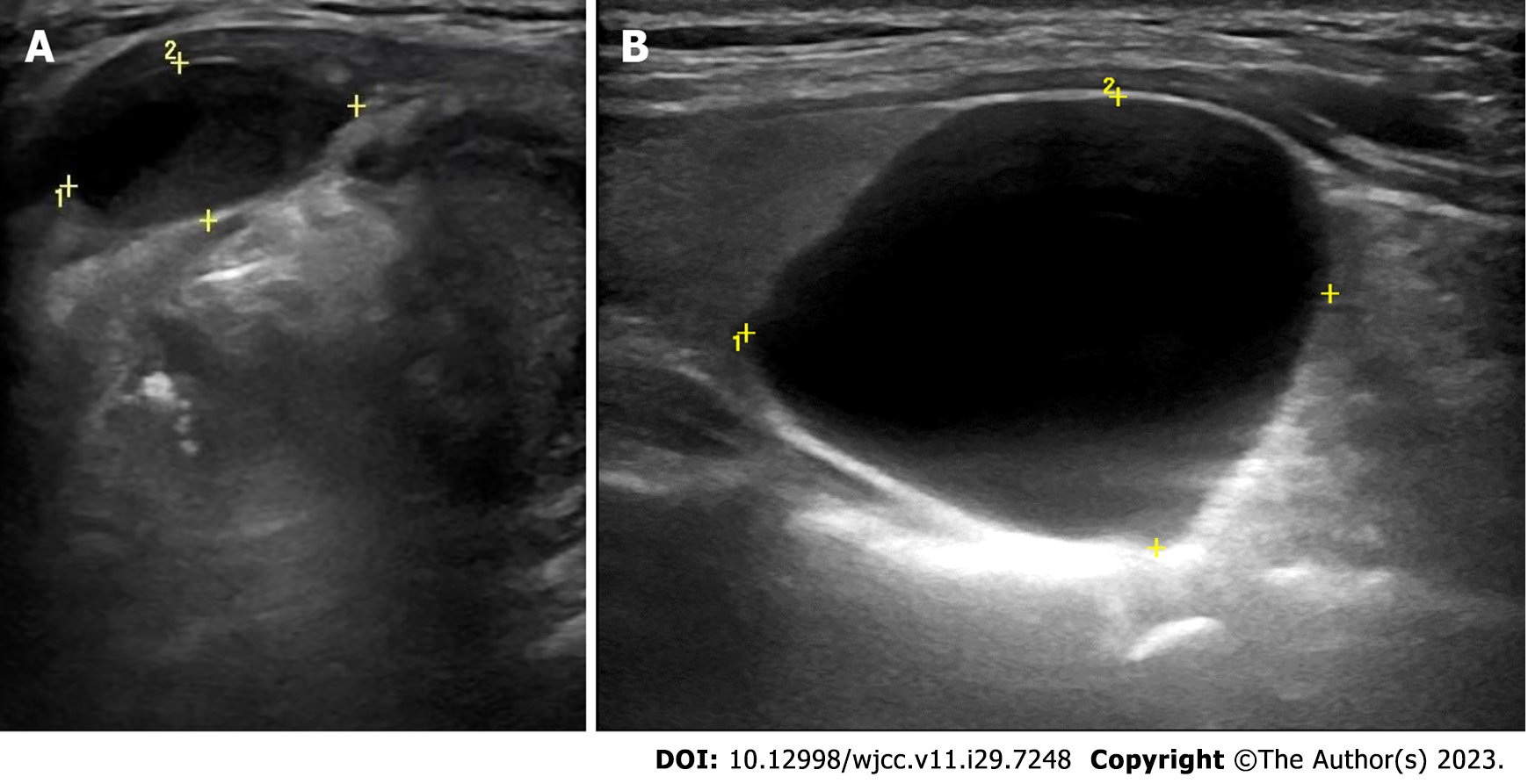
Figure 1 Ultrasonography revealed a thylohyoid cyst and a cystic mass in the left thyroid lobe.
A: The anechoic echo, slightly off-midline to the left, between the thyroid cartilage and strap muscles, without wall inflammation; B: The cystic mass at the lower pole of the left thyroid lobe of the patient.
Figure 2 Computed tomography scan images.
A: The hypodense foci, slightly off-midline to the left, at the left front of the hyoid; B: The lesion at the rear of the lower pole of the left thyroid lobe of the patient.
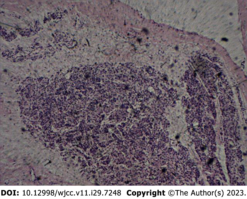
Figure 3
Histochemistry: Cytochemistry immunohistochemistry chromogranin A (+); SynapsinI (-); thyroglobulin (-); thyroid transcription factor-1 (-); cytokeratin 19 antigen (-); nuclear protein Ki-67 about 2%, consistent with parathyroid cyst (hematoxylin and eosin stain, 100×).
- Citation: Chen GY, Li T. Simultaneous thyroglossal duct cyst with parathyroid cyst: A case report. World J Clin Cases 2023; 11(29): 7248-7252
- URL: https://www.wjgnet.com/2307-8960/full/v11/i29/7248.htm
- DOI: https://dx.doi.org/10.12998/wjcc.v11.i29.7248


