Copyright
©The Author(s) 2023.
World J Clin Cases. Oct 16, 2023; 11(29): 7179-7186
Published online Oct 16, 2023. doi: 10.12998/wjcc.v11.i29.7179
Published online Oct 16, 2023. doi: 10.12998/wjcc.v11.i29.7179
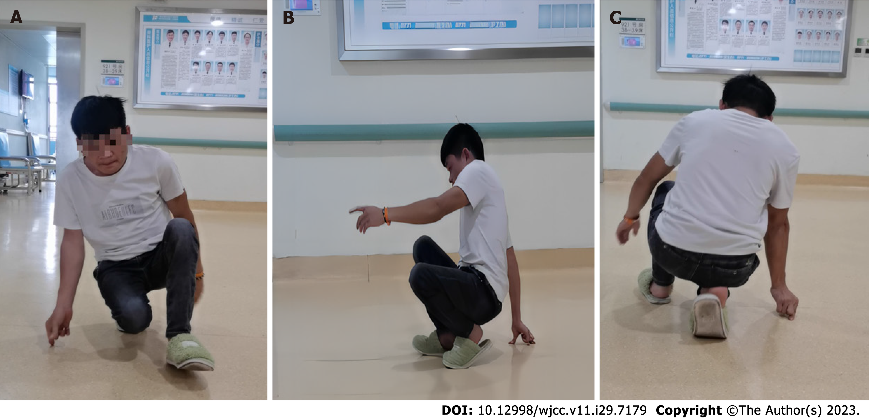
Figure 1 Preoperative squatting gait.
A: Frontal view; B: Lateral view; C: Posterior view.
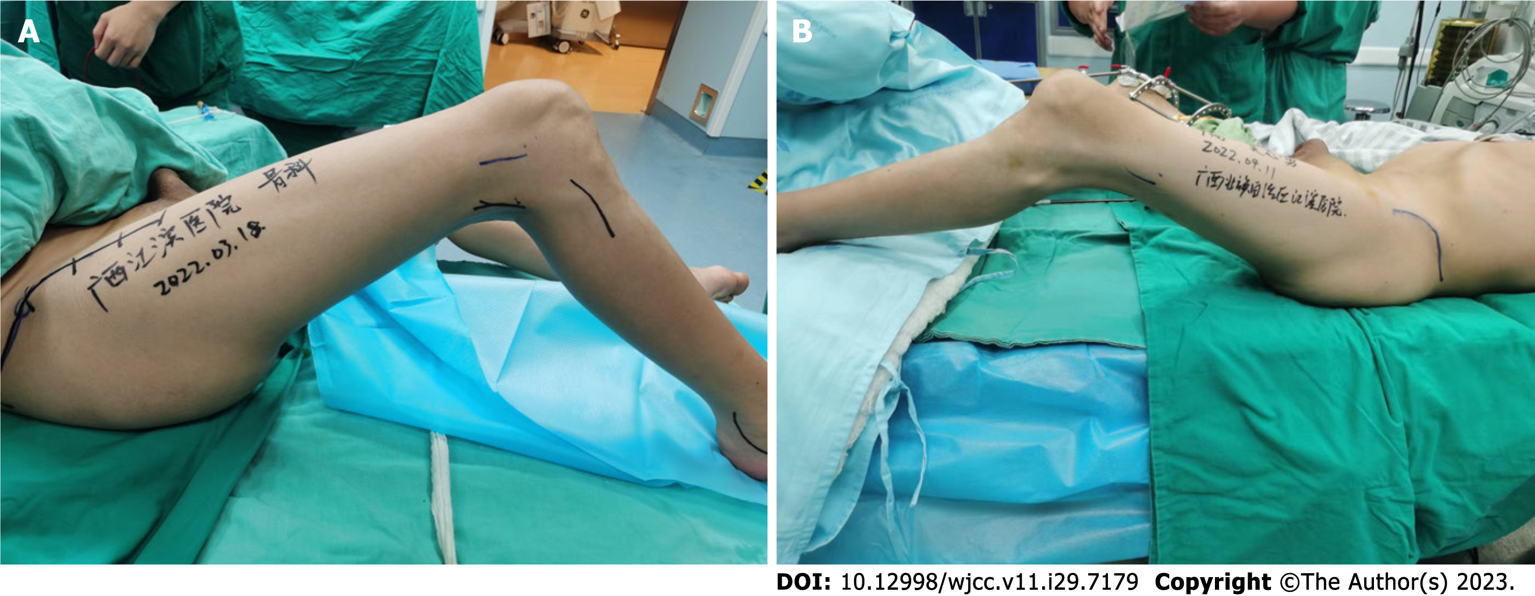
Figure 2 Range of motion of bilateral knees.
A: Range of motion of right knee was 75°-135°; B: Range of motion of left knee was 55°-135°.
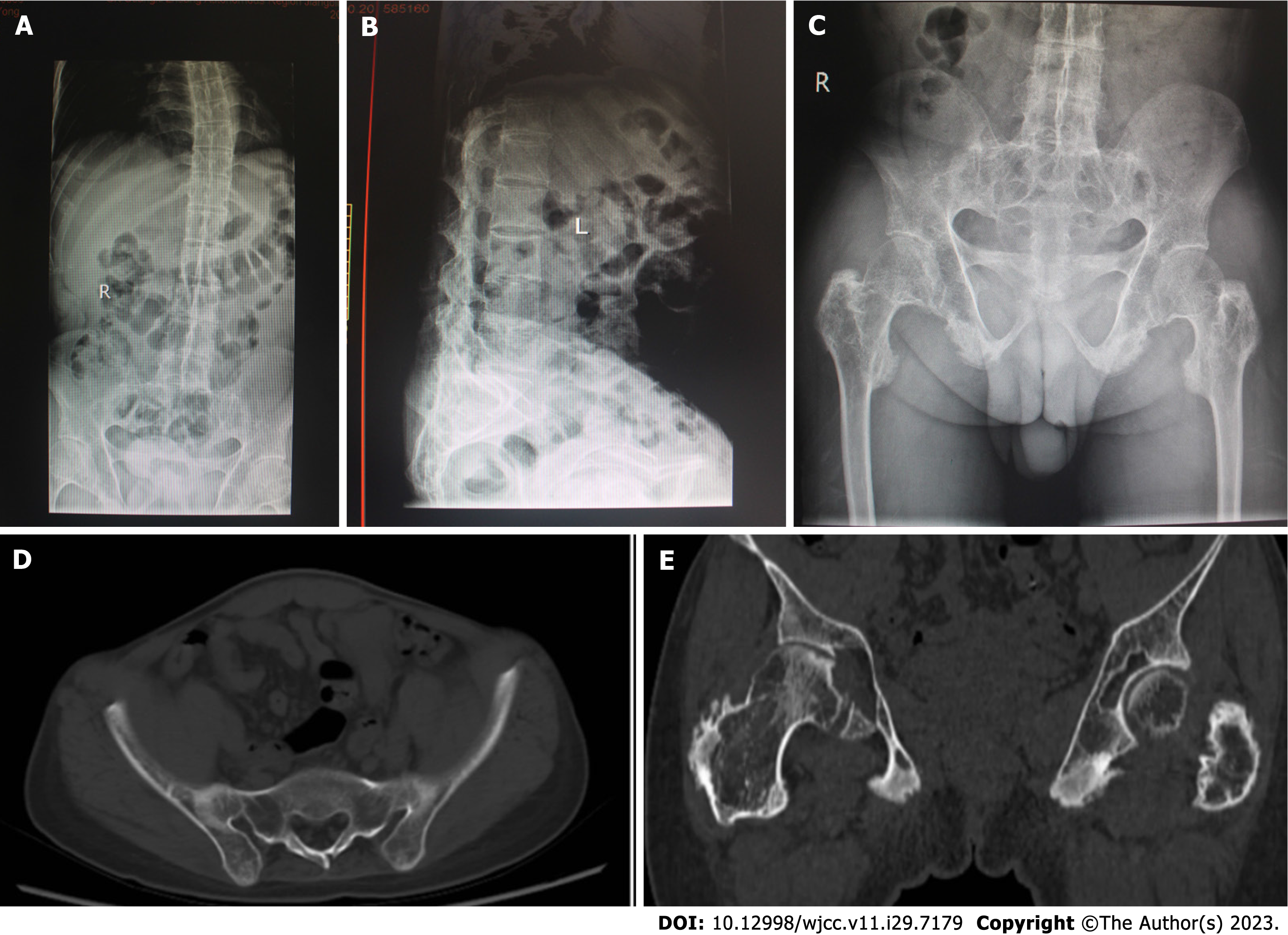
Figure 3 Radiographic changes in the spine, hip joints and sacroiliac joints.
A and B: Plain radiographies of spine showed bamboo spine; C and D: Plain radiography of pelvis and computed tomography scan of pelvis showed sacroiliac joint fusion; E: Computed tomography scan of hips revealed osteoarthritic changes.
Figure 4 Radiographic changes in the knee joints and feet.
A and B: X-rays of the knees revealed osteophytes and bilateral knee flexion contracture; C and D: Plain radiography of ankles showed narrowing of ankle joints space and ankylosing tarsitis.
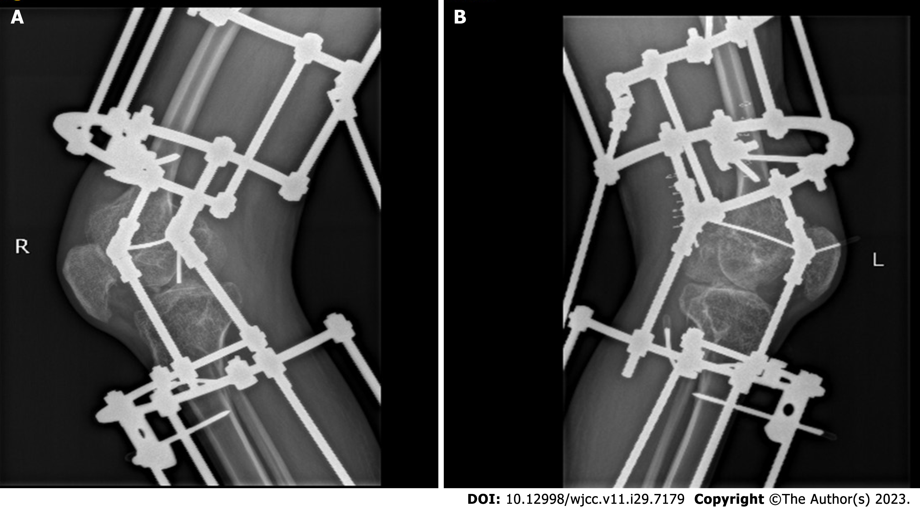
Figure 5 Postoperative X-rays of the bilateral knee joints.
A: The right knee; B: The left knee.
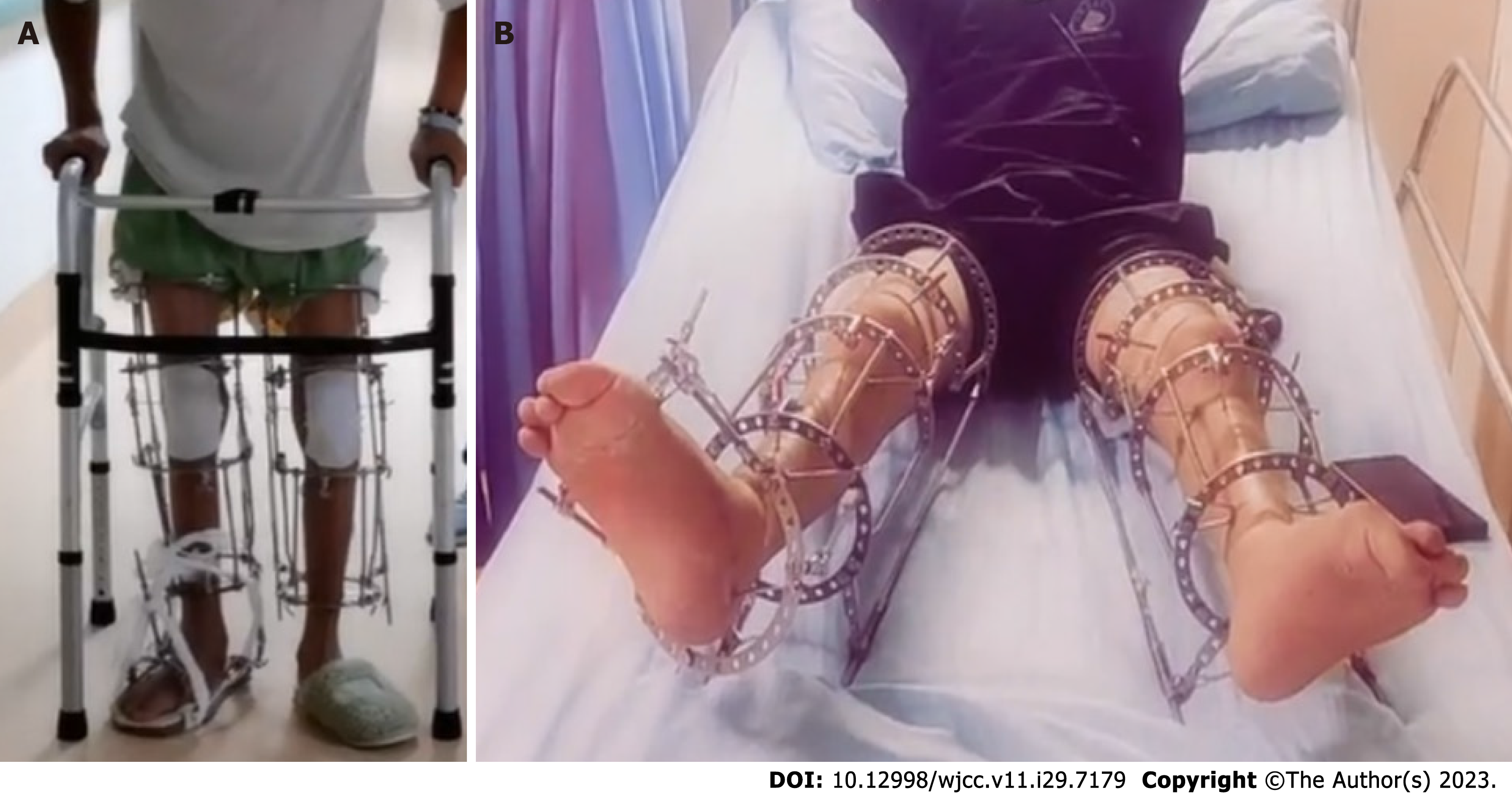
Figure 6 Clinical images before removal of bilateral lower limb ring external fixators.
A: Standing with ring external fixator; B: Showing almost full extension of bilateral knees.
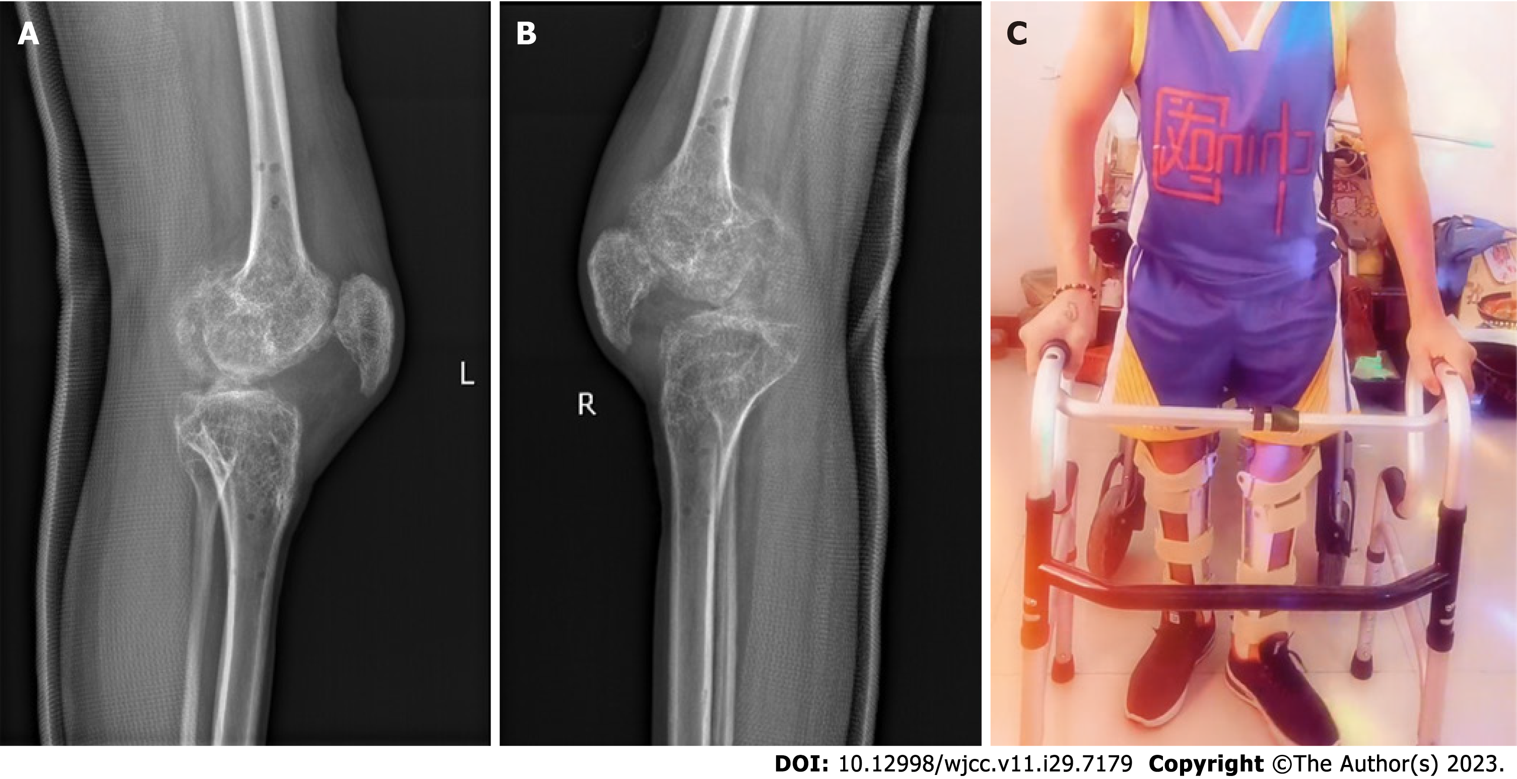
Figure 7 Images after removal of bilateral lower limb ring external fixators.
A and B: Lateral X-rays of bilateral knees; C: Standing up and walking with a walker.
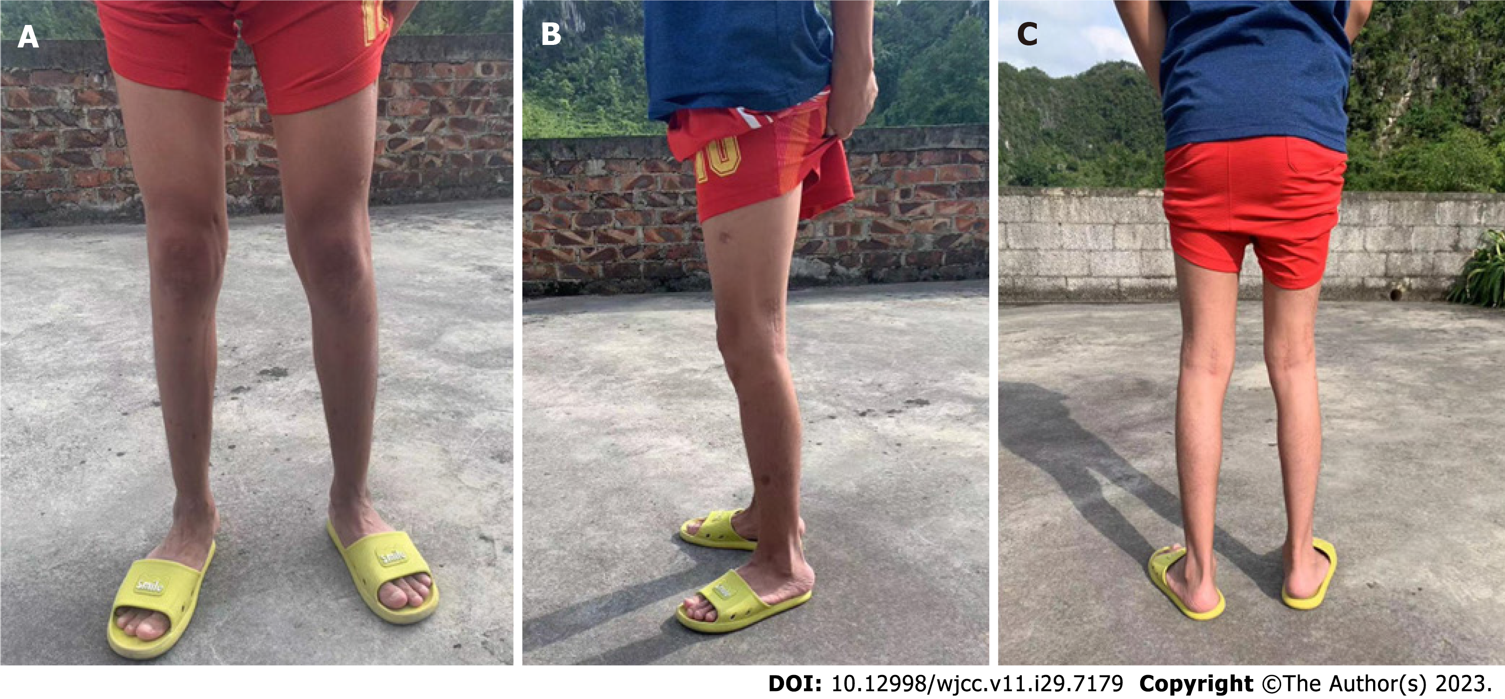
Figure 8 Clinical photographs of the patient after removal of the long-legged brace (12 mo after the operation).
A: Frontal view; B: Lateral view; C: Posterior view.
- Citation: Xia LW, Xu C, Huang JH. Use of Ilizarov technique for bilateral knees flexion contracture in Juvenile-onset ankylosing spondylitis: A case report. World J Clin Cases 2023; 11(29): 7179-7186
- URL: https://www.wjgnet.com/2307-8960/full/v11/i29/7179.htm
- DOI: https://dx.doi.org/10.12998/wjcc.v11.i29.7179


