©The Author(s) 2023.
World J Clin Cases. Sep 16, 2023; 11(26): 6246-6251
Published online Sep 16, 2023. doi: 10.12998/wjcc.v11.i26.6246
Published online Sep 16, 2023. doi: 10.12998/wjcc.v11.i26.6246
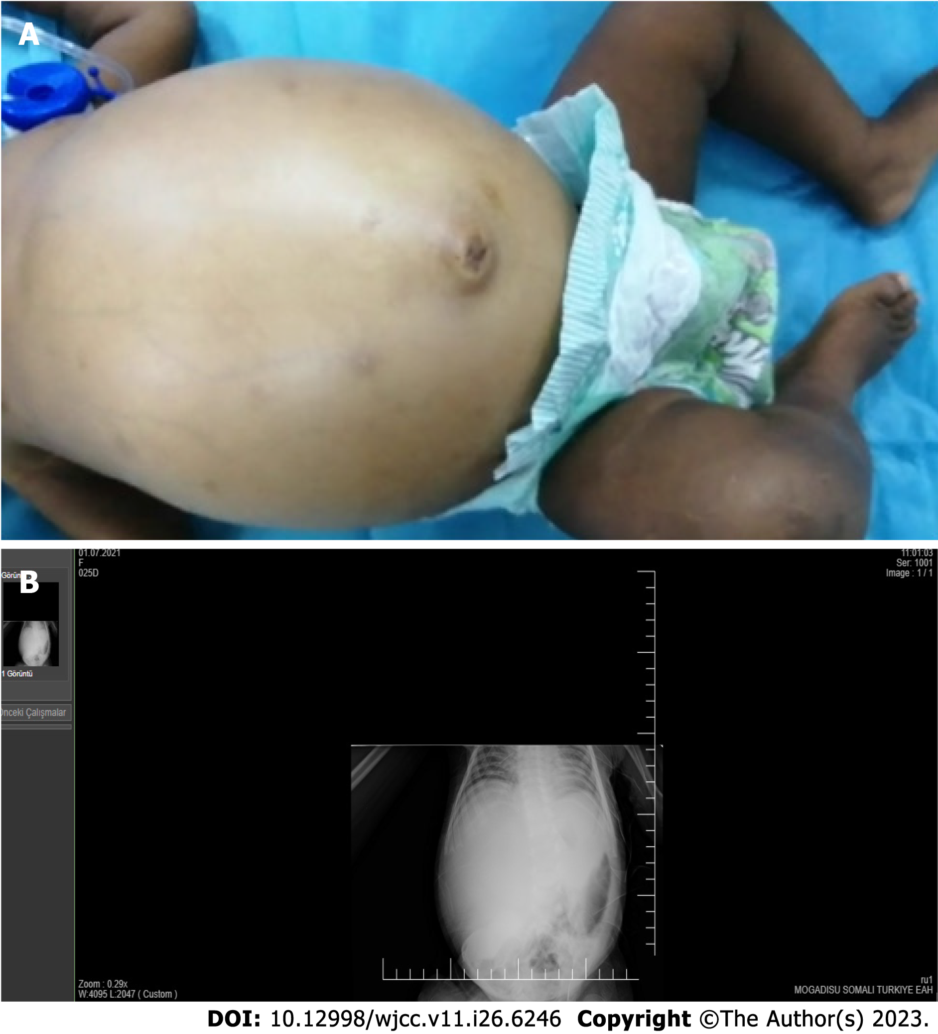
Figure 1 Examination of physical and X-ray.
A: Distension of the abdomen was apparent on physical examination; B: Direct abdominal X-ray.
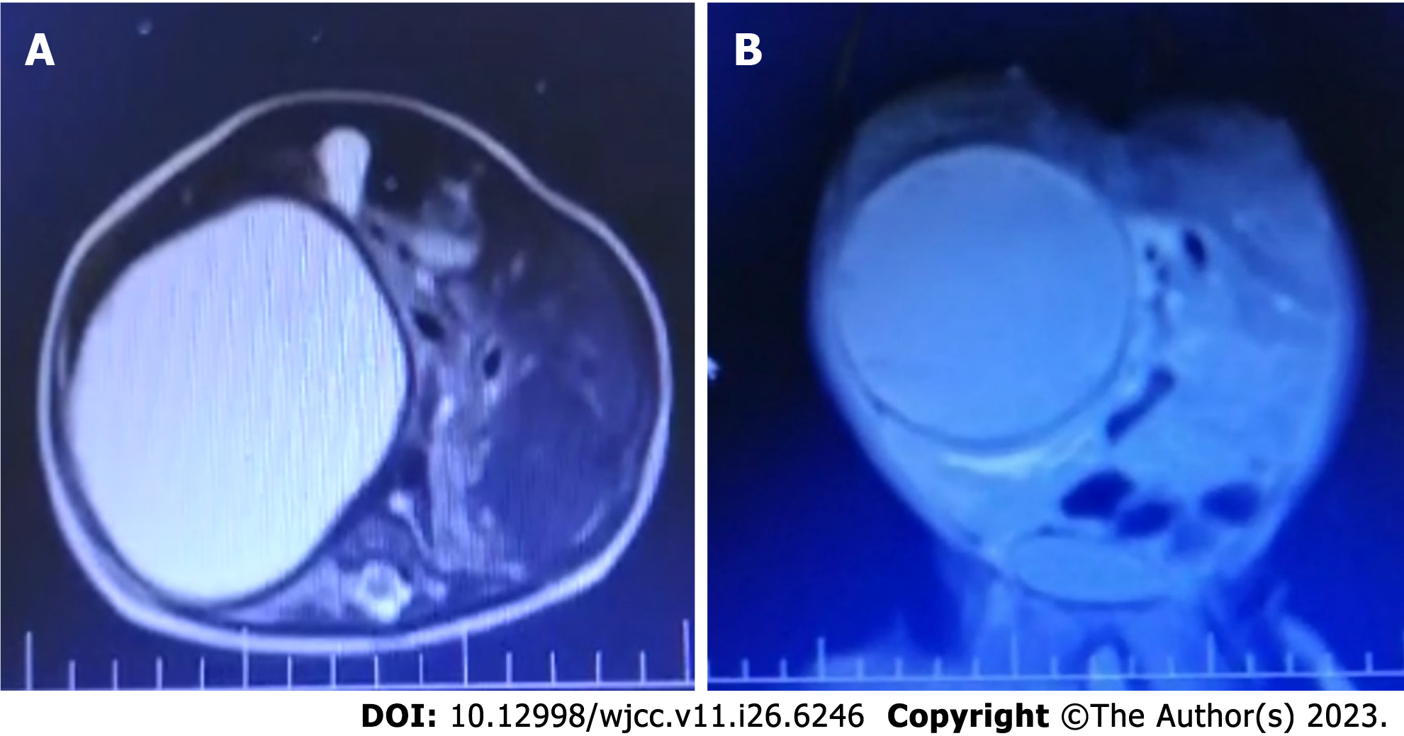
Figure 2 A solitary cystic structure of 10 cm × 10 cm × 14 cm was observed by magnetic resonance imaging in the right abdomen, extending to the pelvis and possibly originating from the liver.
A: Axial image; B: Coronal image.
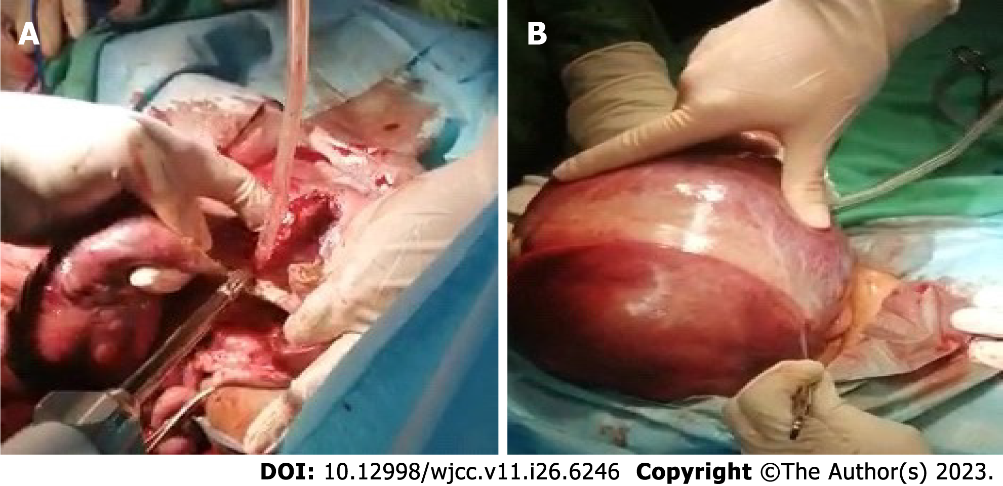
Figure 3 Removal of the cyst adhesions without damaging the vascular structures.
A: Gross appearance of the liver cyst; B: Perioperative view of the excision of the cyst wall.
- Citation: Küçük A, Mohamed SS, Abdi AM, Ali AY. Intestinal obstruction due to giant liver cyst: A case report. World J Clin Cases 2023; 11(26): 6246-6251
- URL: https://www.wjgnet.com/2307-8960/full/v11/i26/6246.htm
- DOI: https://dx.doi.org/10.12998/wjcc.v11.i26.6246


