Copyright
©The Author(s) 2023.
World J Clin Cases. Aug 26, 2023; 11(24): 5721-5728
Published online Aug 26, 2023. doi: 10.12998/wjcc.v11.i24.5721
Published online Aug 26, 2023. doi: 10.12998/wjcc.v11.i24.5721
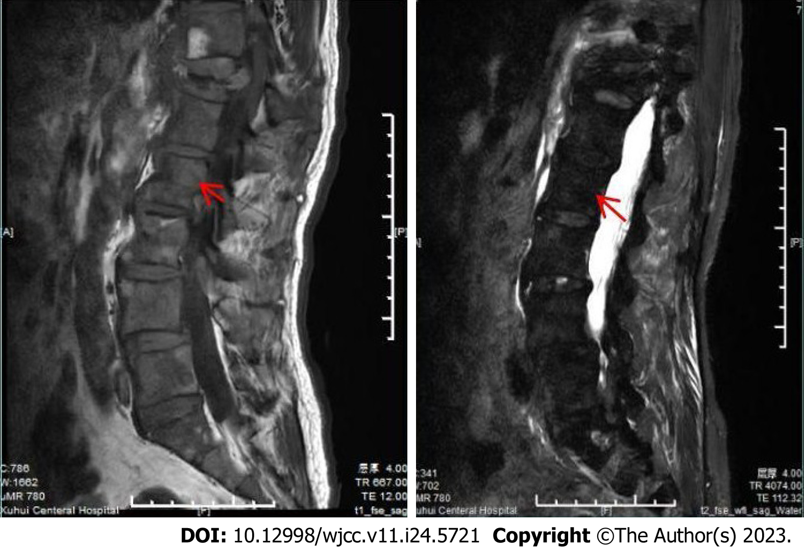
Figure 1 Compressed bone of thoracic 12 vertebrae showing a high T1 signal and low T2 signal.
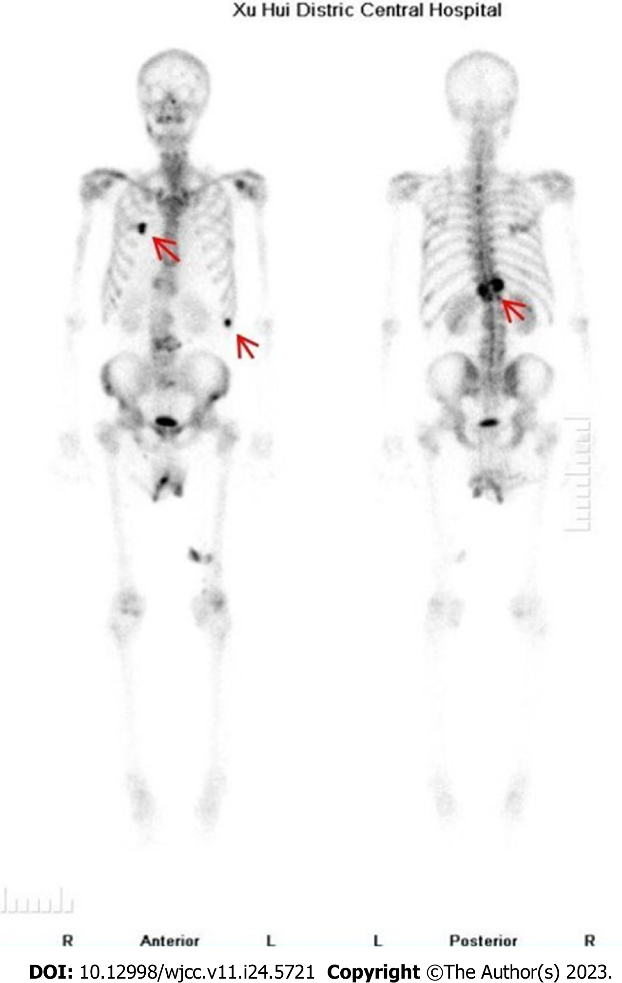
Figure 2 Bone single photon emission tomography/computed tomography showing T12 compression of the whole body bone, and the concentration of radioactivity in bilateral ribs, thus bone metastasis was considered.
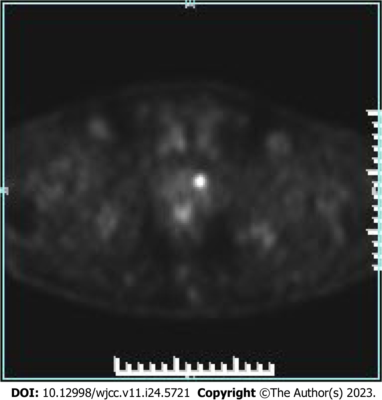
Figure 3 18F-fluorodeoxyglucose positron emission tomography/computed tomography showing focal glucose metabolism in the left lobe of the prostate, and a malignant tumor waiting to be excreted.
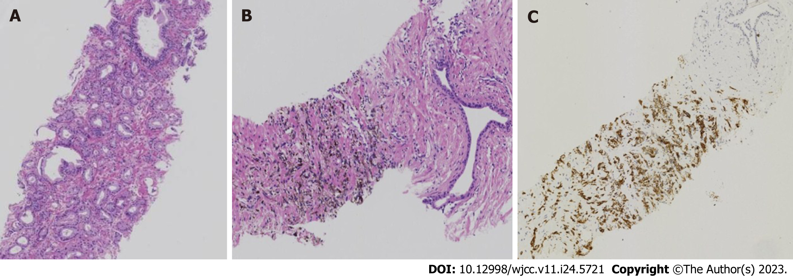
Figure 4 Histopathology.
A: Prostate adenocarcinoma [Hematoxylin and eosin (HE), original magnification, 25 ×]; B: Small foci of melanocytes in the proliferative prostate tissue (HE, original magnification, 25 ×); C: Immunohistochemical results: HMB45+ (HE, original magnification, 25 ×).
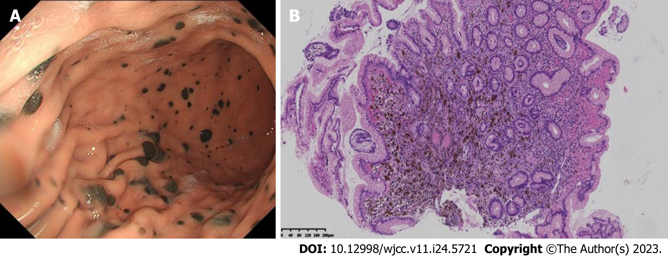
Figure 5 Gastroscopy findings.
A: Multiple mucosal black spots in the body and fundus of the stomach; B: Microscopic small foci of melanocytes in the gastric mucosa (hematoxylin-eosin, original magnification, 25 ×).
- Citation: Zhao H, Liu C, Li B, Guo JM. Malignant melanoma of the prostate: Primary or metastasis? A case report. World J Clin Cases 2023; 11(24): 5721-5728
- URL: https://www.wjgnet.com/2307-8960/full/v11/i24/5721.htm
- DOI: https://dx.doi.org/10.12998/wjcc.v11.i24.5721


