Copyright
©The Author(s) 2023.
World J Clin Cases. Aug 16, 2023; 11(23): 5595-5601
Published online Aug 16, 2023. doi: 10.12998/wjcc.v11.i23.5595
Published online Aug 16, 2023. doi: 10.12998/wjcc.v11.i23.5595
Figure 1 Preoperative plain radiographs of the left knee.
A and B: Plain anteroposterior (A) and lateral (B) radiographs revealed a bony protuberance on the medial side of the patient’s proximal tibia; C and D: Plain anteroposterior (C) and lateral (D) radiographs revealed that osteochondroma was sufficiently resected at immediately after surgery; E and F: Plain anteroposterior (E) and lateral (F) radiographs revealed no recurrence of osteochondroma at one year after surgery.
Figure 2 Preoperative computed tomography scans of the patient’s left knee.
A-C: Axial (A) and 3D (B and C) computed tomography scans showed a bony protuberance with bone marrow continuity on the medial side of the proximal tibia.
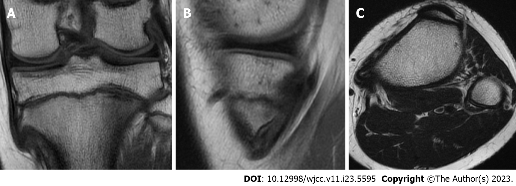
Figure 3 Preoperative T2-weighted magnetic resonance imaging of the left knee.
A-C: Coronal (A), sagittal (B), and axial (C) T2-weighted magnetic resonance imaging of the left knee indicating bone marrow continuity within the lesion and the pes anserinus directly covered the bony protuberance. A high signal change was observed in the semitendinosus tendon.
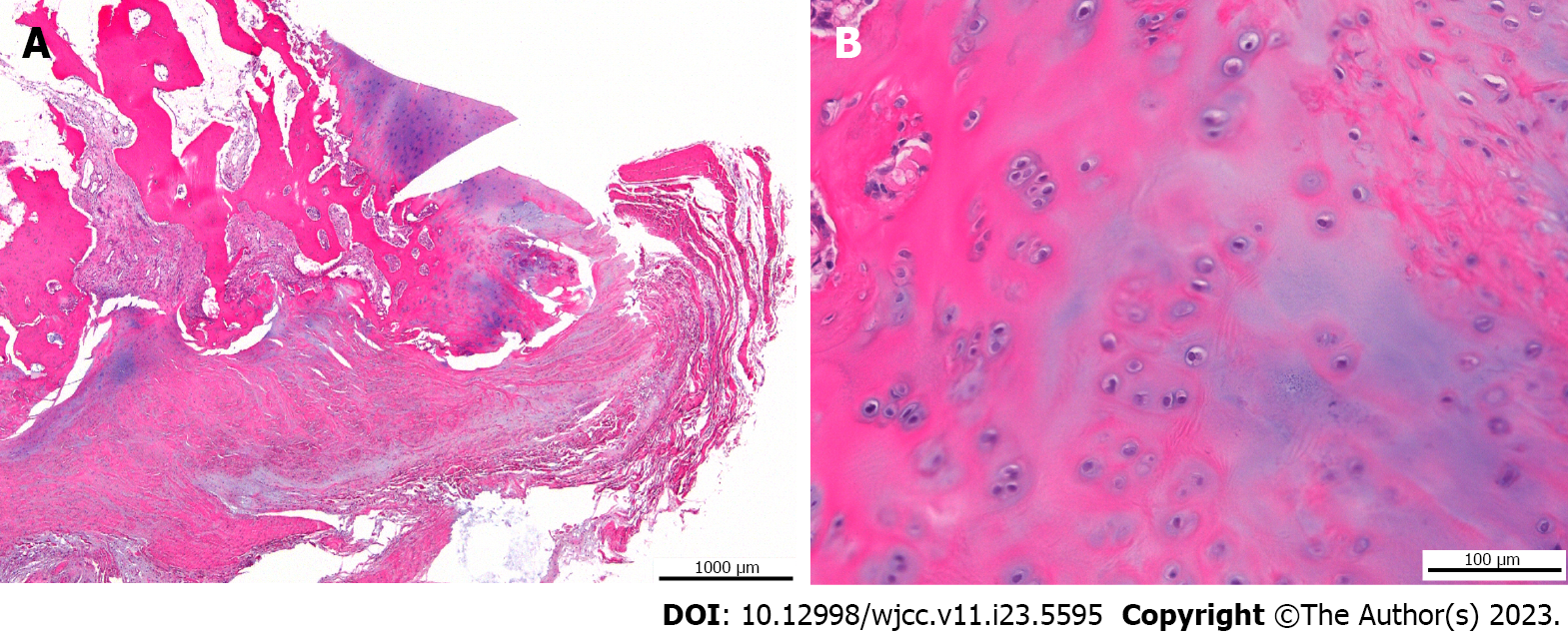
Figure 4 Pathological findings of surgical specimens.
Pathologically, a coating of vitreous cartilage which was thought to be a cartilage cap was observed on the surface of the lesion. A: Hematoxylin-eosin (H-E) staining, × 2; B: H-E staining, × 20.
- Citation: Sonobe T, Hakozaki M, Matsuo Y, Takahashi Y, Yoshida K, Konno S. Knee locking caused by osteochondroma of the proximal tibia adjacent to the pes anserinus: A case report. World J Clin Cases 2023; 11(23): 5595-5601
- URL: https://www.wjgnet.com/2307-8960/full/v11/i23/5595.htm
- DOI: https://dx.doi.org/10.12998/wjcc.v11.i23.5595


