Copyright
©The Author(s) 2023.
World J Clin Cases. Aug 16, 2023; 11(23): 5554-5558
Published online Aug 16, 2023. doi: 10.12998/wjcc.v11.i23.5554
Published online Aug 16, 2023. doi: 10.12998/wjcc.v11.i23.5554
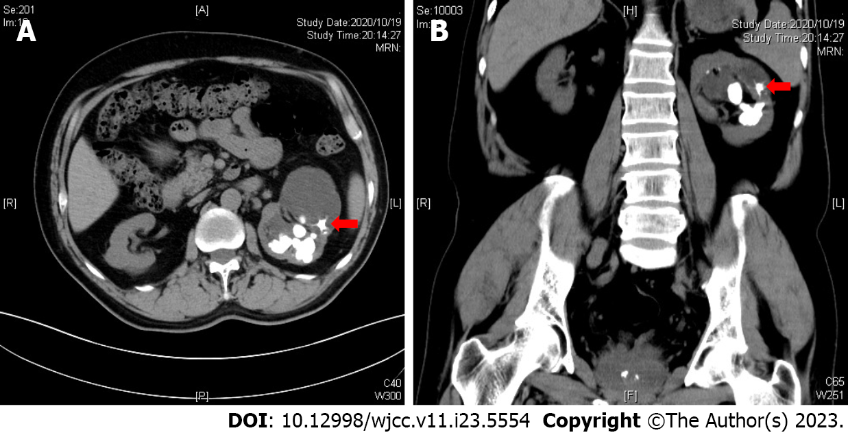
Figure 1 Computed tomography demonstrated a typical jackstone located in the left kidney with hydronephrosis.
A: The axial computed tomography (CT) image. The red arrow points to the jackstone in the renal calyx; B: The coronal CT image. The red arrow points to the jackstone in the renal calyx.
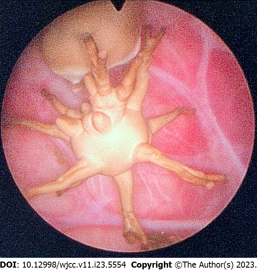
Figure 2 Endoscopic image of the jackstone during percutaneous nephrolithotomy.
It shows that the jackstone with spiky branches is located in an obstructed renal calyx.
- Citation: Song HF, Liang L, Liu YB, Xiao B, Hu WG, Li JX. Jackstone in the renal calyx: A rare case report. World J Clin Cases 2023; 11(23): 5554-5558
- URL: https://www.wjgnet.com/2307-8960/full/v11/i23/5554.htm
- DOI: https://dx.doi.org/10.12998/wjcc.v11.i23.5554


